-
0
Patient Assessment
- 0.1 Patient demand
- 0.2 Overarching considerations
- 0.3 Local history
- 0.4 Anatomical location
- 0.5 General patient history
-
0.6
Risk assessment & special high risk categories
- 5.1 Risk assessment & special high risk categories
- 5.2 age
- 5.3 Compliance
- 5.4 Smoking
- 5.5 Drug abuse
- 5.6 Recreational drugs and alcohol abuse
- 5.7 Parafunctions
- 5.8 Diabetes
- 5.9 Osteoporosis
- 5.10 Coagulation disorders and anticoagulant therapy
- 5.11 Steroids
- 5.12 Bisphosphonates
- 5.13 BRONJ / ARONJ
- 5.14 Radiotherapy
- 5.15 Risk factors
-
1
Diagnostics
-
1.1
Clinical Assessment
- 0.1 Lip line
- 0.2 Mouth opening
- 0.3 Vertical dimension
- 0.4 Maxillo-mandibular relationship
- 0.5 TMD
- 0.6 Existing prosthesis
- 0.7 Muco-gingival junction
- 0.8 Hyposalivation and Xerostomia
- 1.2 Clinical findings
-
1.3
Clinical diagnostic assessments
- 2.1 Microbiology
- 2.2 Salivary output
-
1.4
Diagnostic imaging
- 3.1 Imaging overview
- 3.2 Intraoral radiographs
- 3.3 Panoramic
- 3.4 CBCT
- 3.5 CT
- 1.5 Diagnostic prosthodontic guides
-
1.1
Clinical Assessment
-
2
Treatment Options
- 2.1 Mucosally-supported
-
2.2
Implant-retained/supported, general
- 1.1 Prosthodontic options overview
- 1.2 Number of implants maxilla and mandible
- 1.3 Time to function
- 1.4 Submerged or non-submerged
- 1.5 Soft tissue management
- 1.6 Hard tissue management, mandible
- 1.7 Hard tissue management, maxilla
- 1.8 Need for grafting
- 1.9 Healed vs fresh extraction socket
- 1.10 Digital treatment planning protocols
- 2.3 Implant prosthetics - removable
-
2.4
Implant prosthetics - fixed
- 2.5 Comprehensive treatment concepts
-
3
Treatment Procedures
-
3.1
Surgical
-
3.2
Removable prosthetics
-
3.3
Fixed prosthetics
-
3.1
Surgical
- 4 Aftercare
パノラマX線撮影
Key points
- 医師は認識可能な解剖学的構造に精通していなければなりません。
- 優れたパノラマ写真を獲得するための手順は、テクニックセンシティブです(頭部のポジショニング、顔面/頭部のアクセサリーの取り外し等)。
- デジタルX線撮影では、より多くの選択肢が得られます(ドキュメンテーション、測定、露光ラティチュード)。
パノラマX線撮影
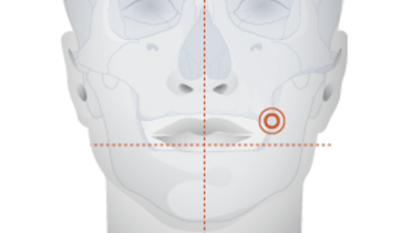
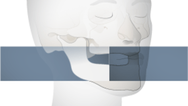
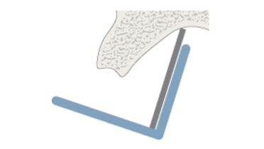
パノラマ写真を撮影する際は、適切な方法に従い、患者の頭蓋をX線ジェネレータとフィルムとの間に注意深くポジショニングすることがきわめて重要です。フィルムと放射線源がスライディングパスに沿って回転するものが多く、放射線照射時間は5~20秒の範囲内です。デジタルX線撮影では、より多くの選択肢があり、より大きな露光ラティチュードが得られます。しかし、パノラマ写真では下顎骨の高さが過小評価され、水平方向の測定値も信頼できません。また、臼歯部では実測値との差がより大きくなります。
パノラマ写真には有効な要素が多数あり、広く用いられています。パノラマ写真は骨内病変や歯を視覚化することができ、上顎洞および眼窩のスクリーニングツールとして用いられています。上顎と下顎の構造にフォーカスするために看過されることの多い頚動脈のアテロームも、パノラマ写真では確認することができます。また、臨床上実用的な利点として、重度の嘔吐反射のある患者に対しても使用することができます。
臨床トピック
Related articles
Additional external resources
Questions
ログインまたはご登録してコメントを投稿してください。
質問する
ログインまたは、無料でご登録して続行してください
You have reached the limit of content accessible without log in or this content requires log in. Log in or sign up now to get unlimited access to all FOR online resources.
FORウェブサイトにご登録していただきますと、すべてのオンライン・リソースに無制限にアクセスできます。FORウェブサイトへのご登録は無料となっております。


