-
0
Patient Assessment
- 0.1 Patient demand
- 0.2 Overarching considerations
- 0.3 Local history
- 0.4 Anatomical location
- 0.5 General patient history
-
0.6
Risk assessment & special high risk categories
- 5.1 Risk assessment & special high risk categories
- 5.2 age
- 5.3 Compliance
- 5.4 Smoking
- 5.5 Drug abuse
- 5.6 Recreational drugs and alcohol abuse
- 5.7 Parafunctions
- 5.8 Diabetes
- 5.9 Osteoporosis
- 5.10 Coagulation disorders and anticoagulant therapy
- 5.11 Steroids
- 5.12 Bisphosphonates
- 5.13 BRONJ / ARONJ
- 5.14 Radiotherapy
- 5.15 Risk factors
-
1
Diagnostics
-
1.1
Clinical Assessment
- 0.1 Lip line
- 0.2 Mouth opening
- 0.3 Vertical dimension
- 0.4 Maxillo-mandibular relationship
- 0.5 TMD
- 0.6 Existing prosthesis
- 0.7 Muco-gingival junction
- 0.8 Hyposalivation and Xerostomia
- 1.2 Clinical findings
-
1.3
Clinical diagnostic assessments
- 2.1 Microbiology
- 2.2 Salivary output
-
1.4
Diagnostic imaging
- 3.1 Imaging overview
- 3.2 Intraoral radiographs
- 3.3 Panoramic
- 3.4 CBCT
- 3.5 CT
- 1.5 Diagnostic prosthodontic guides
-
1.1
Clinical Assessment
-
2
Treatment Options
- 2.1 Mucosally-supported
-
2.2
Implant-retained/supported, general
- 1.1 Prosthodontic options overview
- 1.2 Number of implants maxilla and mandible
- 1.3 Time to function
- 1.4 Submerged or non-submerged
- 1.5 Soft tissue management
- 1.6 Hard tissue management, mandible
- 1.7 Hard tissue management, maxilla
- 1.8 Need for grafting
- 1.9 Healed vs fresh extraction socket
- 1.10 Digital treatment planning protocols
- 2.3 Implant prosthetics - removable
-
2.4
Implant prosthetics - fixed
- 2.5 Comprehensive treatment concepts
-
3
Treatment Procedures
-
3.1
Surgical
-
3.2
Removable prosthetics
-
3.3
Fixed prosthetics
-
3.1
Surgical
- 4 Aftercare
Implant placement with open flap procedure - mandible
Key points
- For overdentures, 2 symphyseal implants placed parallel to the mandibular hinge axis are optimal
- Four implants placed to retain an overdenture are an option
- For fixed prostheses, 4-6 implants installed arch-wise are indicated
- Incisal, mental and mandibular nerves should be protected
- Perforating the lingual cortex in dorsal areas can lead to life-threatening bleeding
Flap preparation - considerations
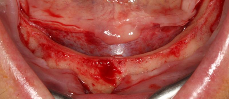
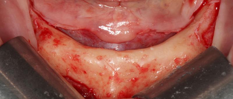
The surgeon should be standing or be seated behind the supine patient. Bilateral truncular anesthesia is needed. The incision can be crestal or at a distance in the labial fold. Presence of keratinized tissue both labially and palatally should ideally be achieved.
The lingual mucoperiosteum is reflected in order to allow for inspection of the mandibular concavities (sublingual, parasymphyseal, submandibular…). Labially the flap should be raised down to the mental foramen. A sagittal release incision reduces the tension of the labial flap.
Osteotomy and implant placement
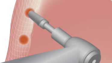
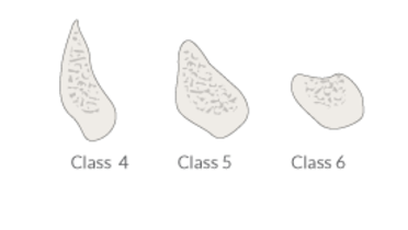
Narrow crests (knife-edge crests) are flattened with a bone forceps to create a surface for pilot drilling. Drilling is performed under ample irrigation with complete in-and-out movements (for evacuation of bone chips). Tactile sensation of the bone density may lead to undersized drilling. Countersinking should be limited to maintain the cortex for stability and resistance remodeling. Primary stability, assessed through recording of the insertion torque and/or Periotest and/or Resonance Frequency Analysis (RFA), will determine the loading protocol - immediate or delayed loading. For implants with medium rough surfaces applying the two-stage protocol means 1 month waiting time.
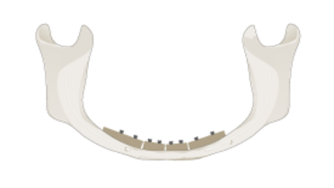
Implants are placed in an arch-wise fashion with an anterio-posterior spread.
Four to six implants are placed anteriorly of the mental foramen. If sufficient bone height above the mandibular canal is present, implant placement can also be located more dorsally. Four implants only will suffice for a fixed cross-arch prosthesis. Placement of 4 implants avoids loading the dorsal mandible by the overdenture. Placement of supplementary implants to deal with an eventual non-integration of implants is not an issue anymore.
To retain an overdenture, 2 implants placed mesially of the mental foramina and parallel to the hinge axis is an optimal solution. In young patients tilting of the overdentures can lead to bone resorption in the dorsal parts of the mandible.
One midline implant only already provides sufficient anchorage for an overdenture.
