-
0
Patient Assessment
- 0.1 Patient demand
- 0.2 Overarching considerations
- 0.3 Local history
- 0.4 Anatomical location
- 0.5 General patient history
-
0.6
Risk assessment & special high risk categories
- 5.1 Risk assessment & special high risk categories
- 5.2 age
- 5.3 Compliance
- 5.4 Smoking
- 5.5 Drug abuse
- 5.6 Recreational drugs and alcohol abuse
- 5.7 Parafunctions
- 5.8 Diabetes
- 5.9 Osteoporosis
- 5.10 Coagulation disorders and anticoagulant therapy
- 5.11 Steroids
- 5.12 Bisphosphonates
- 5.13 BRONJ / ARONJ
- 5.14 Radiotherapy
- 5.15 Risk factors
-
1
Diagnostics
-
1.1
Clinical Assessment
- 0.1 Lip line
- 0.2 Mouth opening
- 0.3 Vertical dimension
- 0.4 Maxillo-mandibular relationship
- 0.5 TMD
- 0.6 Existing prosthesis
- 0.7 Muco-gingival junction
- 0.8 Hyposalivation and Xerostomia
- 1.2 Clinical findings
-
1.3
Clinical diagnostic assessments
- 2.1 Microbiology
- 2.2 Salivary output
-
1.4
Diagnostic imaging
- 3.1 Imaging overview
- 3.2 Intraoral radiographs
- 3.3 Panoramic
- 3.4 CBCT
- 3.5 CT
- 1.5 Diagnostic prosthodontic guides
-
1.1
Clinical Assessment
-
2
Treatment Options
- 2.1 Mucosally-supported
-
2.2
Implant-retained/supported, general
- 1.1 Prosthodontic options overview
- 1.2 Number of implants maxilla and mandible
- 1.3 Time to function
- 1.4 Submerged or non-submerged
- 1.5 Soft tissue management
- 1.6 Hard tissue management, mandible
- 1.7 Hard tissue management, maxilla
- 1.8 Need for grafting
- 1.9 Healed vs fresh extraction socket
- 1.10 Digital treatment planning protocols
- 2.3 Implant prosthetics - removable
-
2.4
Implant prosthetics - fixed
- 2.5 Comprehensive treatment concepts
-
3
Treatment Procedures
-
3.1
Surgical
-
3.2
Removable prosthetics
-
3.3
Fixed prosthetics
-
3.1
Surgical
- 4 Aftercare
Radiological Control
Key points
- Radiological controls are only justified in the presence of certain symptoms: anamnesis and/or clinical examination which demand for further examination
- Digital radiographs are superior, since allowing to send them to (referring) dentist or expert
Intention of radiograph controls
At the beginning of the recall control it is necessary to define the intention of a radiograhical control and if it is needed. Radiological controls are only justified in the presence of certain symptoms, anamnesis and/or clinical examination which require further examination. Such symptoms can be for example clinical signs of peri-implant infection, tissue/bone loss, pain.
Type of radiographs chosen
Depending on what one wants to visualize it can be:
- Intra-oral radiograph, using the paralleling technique, to evaluate the close bone approximation to the implant surface, to measure the marginal bone level around implants, to check the proper abutment-to-implant adaptation and the ventual presence of subgingival cement rests. Implant threads allow precise distance calculations with the shoulder or the top of the implant as reference point. Labial and oral margins do not visualize. A dark line in between the bone and the implant surface does not necessarily mean non-integration but can be due to an optic effect.
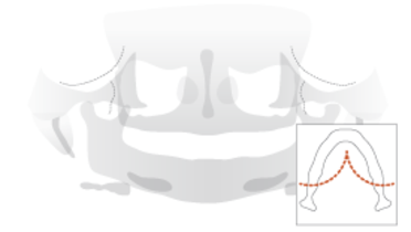
- Panoramic radiograph to have a global picture of the rehabilitated edentulous patient and to detect bone pathologies. One should be aware of distortions inherent to panoramic imaging because of projection geometry. Often in the molar region the magnification factor can reach 1.3. Parallelism of implants cannot be checked on orthopantomograms. Labial and oral margins do not visualize.
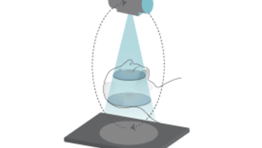
- CBCT may be the method of choice if doubt arises whether an important anatomic structure (mandibular canal …) has been breached. It also allows identifying the labial and oral bone structures. Scatter originating from metallic components can blur the image but software filers are available. CBCT allows evaluating bone augmentation procedures in the oral cavity of in the maxillary sinus.
Clinical topics
Related articles
Questions
Ask a question
Log in or sign up to continue
You have reached the limit of content accessible without log in or this content requires log in. Log in or sign up now to get unlimited access to all FOR online resources.
No payments necessary - FOR is completely free of charge.


