-
0
Patient Assessment
- 0.1 Patient Demand
- 0.2 Anatomical location
-
0.3
Patient History
- 2.1 General patient history
- 2.2 Local history
-
0.4
Risk Assessment
- 3.1 Risk Assessment Overview
- 3.2 Age
- 3.3 Patient Compliance
- 3.4 Smoking
- 3.5 Drug Abuse
- 3.6 Recreational Drug and Alcohol Abuse
- 3.7 Condition of Natural Teeth
- 3.8 Parafunctions
- 3.9 Diabetes
- 3.10 Anticoagulants
- 3.11 Osteoporosis
- 3.12 Bisphosphonates
- 3.13 MRONJ
- 3.14 Steroids
- 3.15 Radiotherapy
- 3.16 Risk factors
-
1
Diagnostics
-
2
Treatment Options
-
2.1
Treatment planning
- 0.1 Non-implant based treatment options
- 0.2 Treatment planning conventional, model based, non-guided, semi-guided
- 0.3 Digital treatment planning
- 0.4 NobelClinician and digital workflow
- 0.5 Implant position considerations overview
- 0.6 Soft tissue condition and morphology
- 0.7 Site development, soft tissue management
- 0.8 Hard tissue and bone quality
- 0.9 Site development, hard tissue management
- 0.10 Time to function
- 0.11 Submerged vs non-submerged
- 0.12 Healed or fresh extraction socket
- 0.13 Screw-retained vs. cement-retained
- 0.14 Angulated Screw Channel system (ASC)
- 2.2 Treatment options esthetic zone
- 2.3 Treatment options posterior zone
- 2.4 Comprehensive treatment concepts
-
2.1
Treatment planning
-
3
Treatment Procedures
-
3.1
Treatment procedures general considerations
- 0.1 Anesthesia
- 0.2 peri-operative care
- 0.3 Flap- or flapless
- 0.4 Non-guided protocol
- 0.5 Semi-guided protocol
- 0.6 Guided protocol overview
- 0.7 Guided protocol NobelGuide
- 0.8 Parallel implant placement considerations
- 0.9 Tapered implant placement considerations
- 0.10 3D implant position
- 0.11 Implant insertion torque
- 0.12 Intra-operative complications
- 0.13 Impression procedures, digital impressions, intraoral scanning
- 3.2 Treatment procedures esthetic zone surgical
- 3.3 Treatment procedures esthetic zone prosthetic
- 3.4 Treatment procedures posterior zone surgical
- 3.5 Treatment procedures posterior zone prosthetic
-
3.1
Treatment procedures general considerations
-
4
Aftercare
治療法の選択肢
Key points
- 単独歯欠損の修復にはいくつかの治療法があり、いずれを選択するかは臨床要因、外科医および歯科医要因ならびに患者要因によって左右されます。
- 単独歯欠損の治療選択肢としては、インプラント支持クラウン、2本の天然歯を支台歯とする3ユニットブリッジ、固定式レジン接着ブリッジ、矯正治療による空隙閉鎖および無治療があります。
治療法の選択肢
単独歯欠損にはいくつかの治療法があり、一般的なのは、インプラント支持クラウン、2本の天然歯を支台歯とする3ユニット固定式ブリッジ、固定式レジン接着ブリッジ、可撤式部分義歯、矯正治療による空隙閉鎖および無治療です。いずれを選択するかは、次のような要因によって左右されます。
- 臨床要因: 残存歯の状態、咬合高径、骨吸収の程度等。
- 外科医および歯科医要因: 治療の実現性に関する知識、各種の治療を遂行するための技能等。
- 患者要因: 最先端の歯科治療へのアクセシビリティ、歯科に関する意識、欠損スペースの位置(前歯部/臼歯部)、(外科的)歯科処置に対する不安、経済的事情等。
インプラント支持補綴物は予後が優れており、患者様の満足度も高く、隣在歯を傷つけずに治療できるという利点があります。しかし、手術に対する不安、欠損スペースの位置、経済的問題等は、インプラント支持クラウンを第一選択としない理由になり得ます。
患者様の希望への対応と誘導
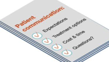
患者様の希望を把握し、適切に対応することは、患者様の満足度を高める最も重要な予測因子です。患者様の声に注意深く耳を傾け、患者様の要求がどのようなものであるかを汲み取り、種々の治療法によって何ができて何ができないかを説明し、これに対して患者様が自らの要望をどのように調整するかを見極める必要があります。治療の実現性について過大な約束をしてはなりません。このため、治療選択肢と各選択肢に関連する重要な要因について患者様に説明する際は、次の点を明らかにする必要があります。
- できること、できないこと
- 予後因子
- 治療期間、治療回数およびタイムライン
- 一時的な苦痛
- 罹患率
- 長期成功率の予後因子とリスク
- 費用および最終的な償還
患者様に提示した種々の治療選択肢の概要をはじめ、治療選択肢の説明を行ったことをカルテに記録し、患者様の反応も記録します。治療法の選択肢は、患者様と一緒に検討する必要があります。各要因の重要度は患者様によってそれぞれ異なり、欠損歯の位置によっても異なります。

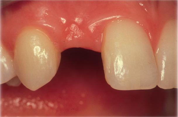
図1: ポジション12(#7 UNIV)の単独歯欠損

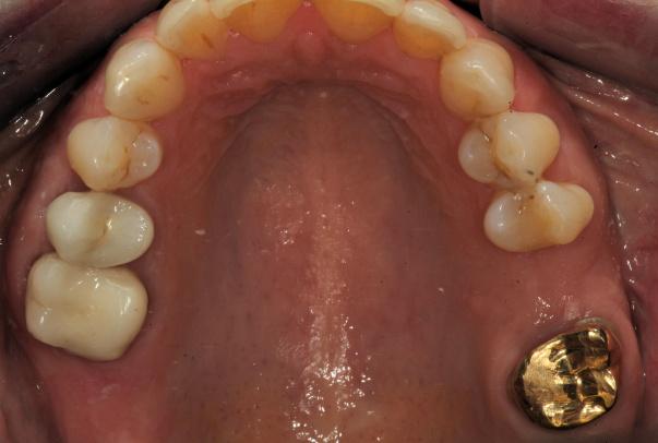
図2: ポジション26(#14 UNIV)の単独歯欠損

