-
0
Patient Assessment
- 0.1 Patient Demand
- 0.2 Anatomical location
-
0.3
Patient History
- 2.1 General patient history
- 2.2 Local history
-
0.4
Risk Assessment
- 3.1 Risk Assessment Overview
- 3.2 Age
- 3.3 Patient Compliance
- 3.4 Smoking
- 3.5 Drug Abuse
- 3.6 Recreational Drug and Alcohol Abuse
- 3.7 Condition of Natural Teeth
- 3.8 Parafunctions
- 3.9 Diabetes
- 3.10 Anticoagulants
- 3.11 Osteoporosis
- 3.12 Bisphosphonates
- 3.13 MRONJ
- 3.14 Steroids
- 3.15 Radiotherapy
- 3.16 Risk factors
-
1
Diagnostics
-
2
Treatment Options
-
2.1
Treatment planning
- 0.1 Non-implant based treatment options
- 0.2 Treatment planning conventional, model based, non-guided, semi-guided
- 0.3 Digital treatment planning
- 0.4 NobelClinician and digital workflow
- 0.5 Implant position considerations overview
- 0.6 Soft tissue condition and morphology
- 0.7 Site development, soft tissue management
- 0.8 Hard tissue and bone quality
- 0.9 Site development, hard tissue management
- 0.10 Time to function
- 0.11 Submerged vs non-submerged
- 0.12 Healed or fresh extraction socket
- 0.13 Screw-retained vs. cement-retained
- 0.14 Angulated Screw Channel system (ASC)
- 2.2 Treatment options esthetic zone
- 2.3 Treatment options posterior zone
- 2.4 Comprehensive treatment concepts
-
2.1
Treatment planning
-
3
Treatment Procedures
-
3.1
Treatment procedures general considerations
- 0.1 Anesthesia
- 0.2 peri-operative care
- 0.3 Flap- or flapless
- 0.4 Non-guided protocol
- 0.5 Semi-guided protocol
- 0.6 Guided protocol overview
- 0.7 Guided protocol NobelGuide
- 0.8 Parallel implant placement considerations
- 0.9 Tapered implant placement considerations
- 0.10 3D implant position
- 0.11 Implant insertion torque
- 0.12 Intra-operative complications
- 0.13 Impression procedures, digital impressions, intraoral scanning
- 3.2 Treatment procedures esthetic zone surgical
- 3.3 Treatment procedures esthetic zone prosthetic
- 3.4 Treatment procedures posterior zone surgical
- 3.5 Treatment procedures posterior zone prosthetic
-
3.1
Treatment procedures general considerations
-
4
Aftercare
診断用模型およびワックスアップ
Key points
- 正確な診断用模型を獲得し、咬合を採得することにより、既存の形態決定因子と治療可能性を高精度で評価することができます。
- 診断用ワックスアップは歯科の診断手法の1つであり、診断用模型の上に装着予定の補綴物をワックスで作製し、患者の希望する審美性と機能を実現するために必要な臨床および技工手順を決定します。
診断用模型およびワックスアップ
インプラント支持補綴物または固定式義歯のいずれも、利用可能なスペースと咬合を診断することが重要です。診断用模型および診断用ワックスアップは、治療の予知性を高めます。
診断用ワックスアップは、単独歯インプラントの診断および治療に使用できるツールの1つで、ワックスまたはワックスにセットした人工歯を使用します。ワックスアップは、ある特定の治療法が適切であるかどうかを示唆する重要な診断情報を提供してくれます。また、適切な補綴方法を選択する手がかりとなり、矯正治療や補綴前手術が必要かどうかを判断することもできます。診断用模型およびワックスアップは、補綴物のために利用できるスペースの量を確認し、補綴用スペースを獲得するために対合歯の治療も必要であるかどうかを確認するのに有効です。また、予定の咬合様式を評価し、残存歯にどのような修正が必要であるかを確認することもできます。加えて、診断用ワックスアップは、歯科医、技工士および患者間のコミュニケーションの手段としても使用することができます。
- 診断用模型およびワックスアップには、以下の手順が含まれます。
- アルジネート印象、咬合採得、必要に応じた写真撮影。
- 2セットの模型作製、咬合器装着(1つは元の状態の保存用、もう1つはワックスアップ用)。
- 診断用ワックスアップ。
- 歯科医、技工士および患者間のコミュニケーション、患者に対する治療計画の3次元的説明。
- 必要に応じて初期計画を調整。
- 治療計画の規模により、口腔内試適を行うため、技工所に常温重合レジンのモックアップ作製を依頼することもできます。
診断用ワックスアップは、治療用ツールとしても使用することができます。無歯顎領域にインプラントを埋入する場合は、これを利用してX線用および外科用のテンプレートを作製することができます。
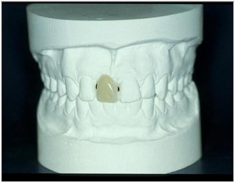
図1:診断用模型とポジション11(#8 UNIV)の欠損歯の診断用ワックスアップ。その他の修復治療は必要ありません。
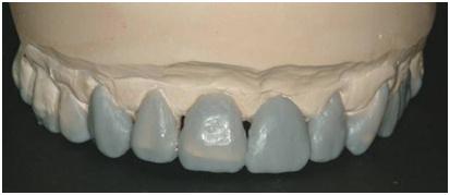
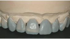
図2:診断用模型とポジション21(#9 UNIV) の欠損歯の診断用ワックスアップ。さらに広範囲の修復治療が必要です。 |


