-
0
Patient Assessment
- 0.1 Patient Demand
- 0.2 Anatomical location
-
0.3
Patient History
- 2.1 General patient history
- 2.2 Local history
-
0.4
Risk Assessment
- 3.1 Risk Assessment Overview
- 3.2 Age
- 3.3 Patient Compliance
- 3.4 Smoking
- 3.5 Drug Abuse
- 3.6 Recreational Drug and Alcohol Abuse
- 3.7 Condition of Natural Teeth
- 3.8 Parafunctions
- 3.9 Diabetes
- 3.10 Anticoagulants
- 3.11 Osteoporosis
- 3.12 Bisphosphonates
- 3.13 MRONJ
- 3.14 Steroids
- 3.15 Radiotherapy
- 3.16 Risk factors
-
1
Diagnostics
-
2
Treatment Options
-
2.1
Treatment planning
- 0.1 Non-implant based treatment options
- 0.2 Treatment planning conventional, model based, non-guided, semi-guided
- 0.3 Digital treatment planning
- 0.4 NobelClinician and digital workflow
- 0.5 Implant position considerations overview
- 0.6 Soft tissue condition and morphology
- 0.7 Site development, soft tissue management
- 0.8 Hard tissue and bone quality
- 0.9 Site development, hard tissue management
- 0.10 Time to function
- 0.11 Submerged vs non-submerged
- 0.12 Healed or fresh extraction socket
- 0.13 Screw-retained vs. cement-retained
- 0.14 Angulated Screw Channel system (ASC)
- 2.2 Treatment options esthetic zone
- 2.3 Treatment options posterior zone
- 2.4 Comprehensive treatment concepts
-
2.1
Treatment planning
-
3
Treatment Procedures
-
3.1
Treatment procedures general considerations
- 0.1 Anesthesia
- 0.2 peri-operative care
- 0.3 Flap- or flapless
- 0.4 Non-guided protocol
- 0.5 Semi-guided protocol
- 0.6 Guided protocol overview
- 0.7 Guided protocol NobelGuide
- 0.8 Parallel implant placement considerations
- 0.9 Tapered implant placement considerations
- 0.10 3D implant position
- 0.11 Implant insertion torque
- 0.12 Intra-operative complications
- 0.13 Impression procedures, digital impressions, intraoral scanning
- 3.2 Treatment procedures esthetic zone surgical
- 3.3 Treatment procedures esthetic zone prosthetic
- 3.4 Treatment procedures posterior zone surgical
- 3.5 Treatment procedures posterior zone prosthetic
-
3.1
Treatment procedures general considerations
-
4
Aftercare
歯周炎
Key points
- 歯周病は、大半の人々がさまざまな形で罹患しています。
- 歯周病は、患者が協力的であれば、適切な治療によって対処することができます。
- 歯周病は、インプラント周囲炎のリスク指標の1つです。
歯周病の特徴
歯周炎は、最も罹患率の高い口腔疾患の1つです。いずれの地域住民でも5~20%が重度の歯周炎に罹患し、成人の大半が軽度ないし中等度の歯周炎に罹患しています。原因因子には局所性と全身性の両方がありますが、歯肉炎や歯周炎のように、通常はプラーク性の炎症が発症因子となります。歯周炎は歯肉炎によって起こると考えられていますが、歯肉炎の罹患部位がすべて歯周炎に進行するとは限りません。通常、歯周炎の最初の徴候が現れるのは、第1大臼歯および切歯です。歯周炎のある患者は、心血管疾患および糖尿病といった種々の全身性疾患の発症リスクも高くなります。
検討事項
単独歯欠損を修復したシングルインプラントの粘膜溝/ポケットには、やがて残存歯の歯肉溝/ポケットと類似した微生物種が認められるようになります。単独歯インプラントの計画作成では、顎全体の状態と全歯の状態を評価します。とりわけ重要なのは、インプラントを埋入する前に歯周病をコントロールし、できれば解消させることです。そのためには口腔衛生指導や衛生士による処置が不可欠であり、ときに歯周手術を行う場合もありますが、何よりも患者の熱心な協力が不可欠です。このような状況下で歯周炎の患者を治療すると、予測実現性の高い良好な転帰が得られます。
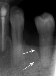
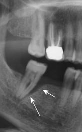
図1-2:下顎左側第2小臼歯および下顎右側第2大臼歯に高度進行歯周病が認められます(矢印)。 この状態でインプラントを埋入することは推奨できません!
歯周病は、喫煙とともに、インプラント周囲にインプラント周囲炎を発症するリスク指標の1つであると考えられており、多数のエビデンスも報告されています。一方で、インプラント治療を受けた患者の絶対多数がインプラント周囲炎の徴候を全く示さないかまたは軽微であり、その多くが現在または過去に歯周炎に罹患していたことから、明確な関係はまだよく分かっていないのが現状です。
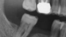
下顎両側のインプラント治療のために紹介された患者。左側第1小臼歯、右側第2小臼歯および両側第2大臼歯の抜去を計画しています。
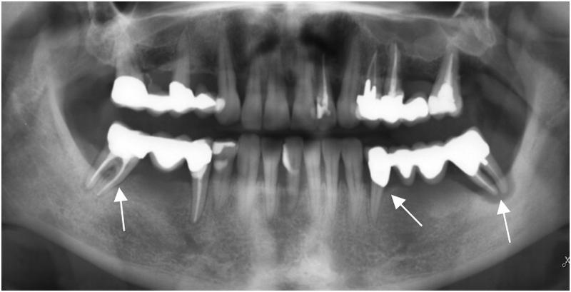
図3: 複数の歯に高度進行歯周炎が認められます(白色矢印)。
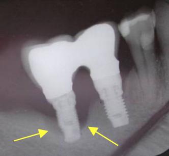
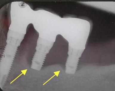
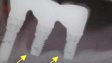
図4および図5:埋入インプラント5本のうち3本が埋入後3~4年で重度のインプラント周囲炎を発症し(黄色矢印)、歯周病に似た外観を呈しています。

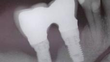
図6: 埋入5年後、非罹患インプラントが左右に1本ずつ残存しています。
