-
0
Patient Assessment
- 0.1 Patient Demand
- 0.2 Anatomical location
-
0.3
Patient History
- 2.1 General patient history
- 2.2 Local history
-
0.4
Risk Assessment
- 3.1 Risk Assessment Overview
- 3.2 Age
- 3.3 Patient Compliance
- 3.4 Smoking
- 3.5 Drug Abuse
- 3.6 Recreational Drug and Alcohol Abuse
- 3.7 Condition of Natural Teeth
- 3.8 Parafunctions
- 3.9 Diabetes
- 3.10 Anticoagulants
- 3.11 Osteoporosis
- 3.12 Bisphosphonates
- 3.13 MRONJ
- 3.14 Steroids
- 3.15 Radiotherapy
- 3.16 Risk factors
-
1
Diagnostics
-
2
Treatment Options
-
2.1
Treatment planning
- 0.1 Non-implant based treatment options
- 0.2 Treatment planning conventional, model based, non-guided, semi-guided
- 0.3 Digital treatment planning
- 0.4 NobelClinician and digital workflow
- 0.5 Implant position considerations overview
- 0.6 Soft tissue condition and morphology
- 0.7 Site development, soft tissue management
- 0.8 Hard tissue and bone quality
- 0.9 Site development, hard tissue management
- 0.10 Time to function
- 0.11 Submerged vs non-submerged
- 0.12 Healed or fresh extraction socket
- 0.13 Screw-retained vs. cement-retained
- 0.14 Angulated Screw Channel system (ASC)
- 2.2 Treatment options esthetic zone
- 2.3 Treatment options posterior zone
- 2.4 Comprehensive treatment concepts
-
2.1
Treatment planning
-
3
Treatment Procedures
-
3.1
Treatment procedures general considerations
- 0.1 Anesthesia
- 0.2 peri-operative care
- 0.3 Flap- or flapless
- 0.4 Non-guided protocol
- 0.5 Semi-guided protocol
- 0.6 Guided protocol overview
- 0.7 Guided protocol NobelGuide
- 0.8 Parallel implant placement considerations
- 0.9 Tapered implant placement considerations
- 0.10 3D implant position
- 0.11 Implant insertion torque
- 0.12 Intra-operative complications
- 0.13 Impression procedures, digital impressions, intraoral scanning
- 3.2 Treatment procedures esthetic zone surgical
- 3.3 Treatment procedures esthetic zone prosthetic
- 3.4 Treatment procedures posterior zone surgical
- 3.5 Treatment procedures posterior zone prosthetic
-
3.1
Treatment procedures general considerations
-
4
Aftercare
模型ベース、ノンガイデッド、セミガイデッドプランニング
Key points
- インプラントを骨の適切な位置に埋入するため、診断用のプロビジョナルセットアップを利用して、サージカルテンプレートを作製することもできます。
- サージカルテンプレートの作製には、従来型の模型を用いる方法とコーンビームCTを用いる方法があります。
サージカルテンプレートは、手術時にインプラントを適切な位置に埋入するための補助具として使用されます。サージカルテンプレートによって提供される外科的制約の度合いにより、次の3つのデザインコンセプトがあります。
- ノンリミッティングデザイン
- セミガイデッドデザイン:埋入窩形成に使用する最初のドリルはサージカルテンプレートを利用して方向を定めます。
- フルガイデッドデザイン
ノンリミッティングデザインおよびセミガイデッドデザインでは、従来型の模型を用いますが、フルガイデッドデザインでは、コーンビームCTとプランニングソフトウェアを用います。
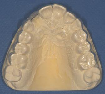
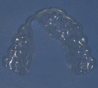
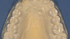
ノンリミッティングデザインは、模型とワックスアップに基づいて真空成型のテンプレートを作製し、補綴物の咬合部にオープンスペースを設けます。このデザインは埋入角度を指示せず、選択されたインプラント埋入部位に対して装着予定の補綴物がどこに位置するかを示す指標のみを提示します(図1および2)。
セミガイデッドデザインも、模型とワックスアップに基づき、インプラント支持補綴物の装着予定位置を含む残存歯列の咬合部上にアクリリックレジンでマトリックスを作製します。装着予定の補綴物に対して理想的なインプラントの埋入位置および角度に合わせ、アクリリックレジンに孔を開けます。唇側または口蓋/舌側のアクリリックレジンを取り除き、最初のドリルの位置および角度のためにhalf-openのチューブを残します。サージカルテンプレートを用いて埋入窩形成に使用する最初のドリルの方向を定め、残りの埋入窩形成とインプラント埋入はフリーハンドで行うため、一定の柔軟性が残されています。
Digital Textbooks
Achieving your intended outcome with endosseous implant therapy requires careful planning and execution of both the surgical and prosthodontic aspects of treatment. Seamless integration of these two phases of care is vital. Attaining your final treatment outcome typically is based on a number of factors: a comprehensive evaluation of the patient, obtaining the required records, a thorough understanding of the possibilities and limitations inherent in both the surgical and prosthodontic phases of different treatment options, developing and communicating a definitive treatment plan to the patient, and patient acceptance of that treatment plan.
Proper surgical planning requires that the clinicians involved in the care of the patient jointly discuss the various phases of the patient’s treatment, beginning with a thorough diagnosis and detailed treatment plan. The restoring dentist and the surgeon, using a team approach, must decide on all the treatment details prior to the surgical appointment. The team first must decide if the patient is an appropriate candidate for a dental implant. Once it is determined the patient is suitable, the edentulous ridge should be analyzed to determine if there is sufficient bone present to permit implant placement. If so, then the precise position and form of the crown should be identified. The position of that crown can be transferred to a cone beam computed tomography (CBCT) scan for the purpose of fabricating a surgical template to guide implant placement precisely during the actual surgery.
Questions
ログインまたはご登録してコメントを投稿してください。
質問する
ログインまたは、無料でご登録して続行してください
You have reached the limit of content accessible without log in or this content requires log in. Log in or sign up now to get unlimited access to all FOR online resources.
FORウェブサイトにご登録していただきますと、すべてのオンライン・リソースに無制限にアクセスできます。FORウェブサイトへのご登録は無料となっております。


