-
0
Patient Assessment
- 0.1 Patient Demand
- 0.2 Anatomical location
-
0.3
Patient History
- 2.1 General patient history
- 2.2 Local history
-
0.4
Risk Assessment
- 3.1 Risk Assessment Overview
- 3.2 Age
- 3.3 Patient Compliance
- 3.4 Smoking
- 3.5 Drug Abuse
- 3.6 Recreational Drug and Alcohol Abuse
- 3.7 Condition of Natural Teeth
- 3.8 Parafunctions
- 3.9 Diabetes
- 3.10 Anticoagulants
- 3.11 Osteoporosis
- 3.12 Bisphosphonates
- 3.13 MRONJ
- 3.14 Steroids
- 3.15 Radiotherapy
- 3.16 Risk factors
-
1
Diagnostics
-
2
Treatment Options
-
2.1
Treatment planning
- 0.1 Non-implant based treatment options
- 0.2 Treatment planning conventional, model based, non-guided, semi-guided
- 0.3 Digital treatment planning
- 0.4 NobelClinician and digital workflow
- 0.5 Implant position considerations overview
- 0.6 Soft tissue condition and morphology
- 0.7 Site development, soft tissue management
- 0.8 Hard tissue and bone quality
- 0.9 Site development, hard tissue management
- 0.10 Time to function
- 0.11 Submerged vs non-submerged
- 0.12 Healed or fresh extraction socket
- 0.13 Screw-retained vs. cement-retained
- 0.14 Angulated Screw Channel system (ASC)
- 2.2 Treatment options esthetic zone
- 2.3 Treatment options posterior zone
- 2.4 Comprehensive treatment concepts
-
2.1
Treatment planning
-
3
Treatment Procedures
-
3.1
Treatment procedures general considerations
- 0.1 Anesthesia
- 0.2 peri-operative care
- 0.3 Flap- or flapless
- 0.4 Non-guided protocol
- 0.5 Semi-guided protocol
- 0.6 Guided protocol overview
- 0.7 Guided protocol NobelGuide
- 0.8 Parallel implant placement considerations
- 0.9 Tapered implant placement considerations
- 0.10 3D implant position
- 0.11 Implant insertion torque
- 0.12 Intra-operative complications
- 0.13 Impression procedures, digital impressions, intraoral scanning
- 3.2 Treatment procedures esthetic zone surgical
- 3.3 Treatment procedures esthetic zone prosthetic
- 3.4 Treatment procedures posterior zone surgical
- 3.5 Treatment procedures posterior zone prosthetic
-
3.1
Treatment procedures general considerations
-
4
Aftercare
補綴診断用ツールの概要
Key points
- 単独歯補綴に利用できる3次元的スペースの測定は、治療前診断の重要な項目の1つです。
- 正確な診断用模型を獲得し、咬合/歯列弓を採得することにより、既存の形態決定因子と治療可能性を高精度で評価することができます。
概要
単独歯のインプラント支持補綴が関与するのは歯列のごくわずかな部分ですが、利用可能なスペースと咬合を診断することは、きわめて重要です。単独歯補綴と咬合に利用できる3次元的スペースを測定するため、専用ツールを利用することもできます。
方法は以下のとおりです。
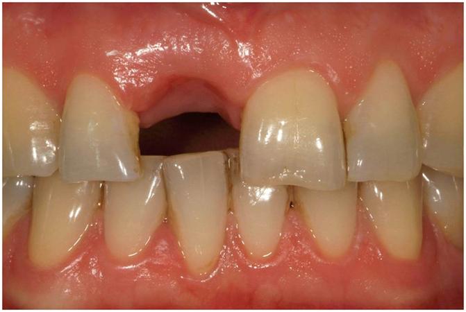
- 対合歯と目視で比較することにより、適切な修復スペースを確認します。対合歯が正常な幅を有する場合は、これを指標として欠損歯修復のための十分なスペースを決定することができます。
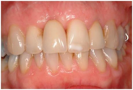
- 暫間補綴物のサイズを目視で検査し、暫間補綴物が抜歯空隙を違和感なく満たしているかどうかを確認します。
- 口腔内光学スキャナーを用いて歯列弓と咬合関係のコンピュータ模型を獲得します。診断のため、ソフトウェアプログラムで人工歯を追加することもできます。
- 石膏模型を作製し、咬合採得を行います。従来型の診断用ワックスアップを行い、利用可能なスペースを検討します。
- 石膏模型を作製し、咬合採得を行います。歯科技工所で光学スキャナーを用いて模型を読み込むことにより、コンピュータ模型を作製し、ソフトウェアプログラムで人工歯を追加することができます。
コーンビームCTと併せて上述の方法を実践することにより、補綴物のために利用できるスペース、インプラント埋入に利用できる骨の量や形状、周囲の解剖学的構造との位置関係について十分なデータを獲得することができます。また、各診断方法の費用効果も念頭に置く必要があります。大がかりな方法は費用もかかるため、すべての症例に用いる必要はありません。
図1:ポジション11(#8 UNIV)にインプラント支持補綴物を希望している患者。目視で対合歯と比較することにより適切な補綴スペースを決定する方法。
図2:ポジション11(#8 UNIV)にインプラント支持補綴物を希望している患者。暫間補綴物のサイズを決定するために目視で確認し、抜歯空隙を違和感なく満たす方法。
Digital Textbooks
Achieving your intended outcome with endosseous implant therapy requires careful planning and execution of both the surgical and prosthodontic aspects of treatment. Seamless integration of these two phases of care is vital. Attaining your final treatment outcome typically is based on a number of factors: a comprehensive evaluation of the patient, obtaining the required records, a thorough understanding of the possibilities and limitations inherent in both the surgical and prosthodontic phases of different treatment options, developing and communicating a definitive treatment plan to the patient, and patient acceptance of that treatment plan.
Questions
ログインまたはご登録してコメントを投稿してください。
質問する
ログインまたは、無料でご登録して続行してください
You have reached the limit of content accessible without log in or this content requires log in. Log in or sign up now to get unlimited access to all FOR online resources.
FORウェブサイトにご登録していただきますと、すべてのオンライン・リソースに無制限にアクセスできます。FORウェブサイトへのご登録は無料となっております。


Recent diagnostic tools in fixed prosthodontics