-
0
Patient Assessment
- 0.1 Patient Demand
- 0.2 Anatomical location
-
0.3
Patient History
- 2.1 General patient history
- 2.2 Local history
-
0.4
Risk Assessment
- 3.1 Risk Assessment Overview
- 3.2 Age
- 3.3 Patient Compliance
- 3.4 Smoking
- 3.5 Drug Abuse
- 3.6 Recreational Drug and Alcohol Abuse
- 3.7 Condition of Natural Teeth
- 3.8 Parafunctions
- 3.9 Diabetes
- 3.10 Anticoagulants
- 3.11 Osteoporosis
- 3.12 Bisphosphonates
- 3.13 MRONJ
- 3.14 Steroids
- 3.15 Radiotherapy
- 3.16 Risk factors
-
1
Diagnostics
-
2
Treatment Options
-
2.1
Treatment planning
- 0.1 Non-implant based treatment options
- 0.2 Treatment planning conventional, model based, non-guided, semi-guided
- 0.3 Digital treatment planning
- 0.4 NobelClinician and digital workflow
- 0.5 Implant position considerations overview
- 0.6 Soft tissue condition and morphology
- 0.7 Site development, soft tissue management
- 0.8 Hard tissue and bone quality
- 0.9 Site development, hard tissue management
- 0.10 Time to function
- 0.11 Submerged vs non-submerged
- 0.12 Healed or fresh extraction socket
- 0.13 Screw-retained vs. cement-retained
- 0.14 Angulated Screw Channel system (ASC)
- 2.2 Treatment options esthetic zone
- 2.3 Treatment options posterior zone
- 2.4 Comprehensive treatment concepts
-
2.1
Treatment planning
-
3
Treatment Procedures
-
3.1
Treatment procedures general considerations
- 0.1 Anesthesia
- 0.2 peri-operative care
- 0.3 Flap- or flapless
- 0.4 Non-guided protocol
- 0.5 Semi-guided protocol
- 0.6 Guided protocol overview
- 0.7 Guided protocol NobelGuide
- 0.8 Parallel implant placement considerations
- 0.9 Tapered implant placement considerations
- 0.10 3D implant position
- 0.11 Implant insertion torque
- 0.12 Intra-operative complications
- 0.13 Impression procedures, digital impressions, intraoral scanning
- 3.2 Treatment procedures esthetic zone surgical
- 3.3 Treatment procedures esthetic zone prosthetic
- 3.4 Treatment procedures posterior zone surgical
- 3.5 Treatment procedures posterior zone prosthetic
-
3.1
Treatment procedures general considerations
-
4
Aftercare
テーパード・インプラント埋入に関する検討事項
Key points
- テーパード・インプラントのデザイン=歯根形状。
- 適応:インプラントの即時埋入、あらゆる種類の骨質、特に軟らかい骨質、隣接歯の歯根が近接し間隙が狭い場合。
- 適切な埋入トルクと初期固定。
- 今日、最も多く使用されているインプラントのデザイン。
長所
今日、多く使用されているインプラントは、歯根の形状に類似したテーパー状のデザインです。ストレートウォールと比較した場合のテーパー形状の主な長所は、インプラントの即時埋入に理想的であること、隣接歯の歯根が近接した狭い間隙用にうまく設計されていること、スクリューデザインにより初期固定が良好であること、軟らかい骨質、先端の径が全方向に対し細くなっているために穿孔リスクが低いことです。テーパード・インプラントはあらゆる骨質に適応となりますが、骨を局所的に圧縮し、その結果インプラントの固定を高めるため、軟らかい骨質(D4)において際立った長所があります。この長所は、トランスクレスタルの上顎洞拳上術等、増生/移植を同時に行う手術で活用できます。
製品の選択肢
ノーベルバイオケアのテーパード・インプラントデザインには、ノーベルテーパード・コニカル・コネクション(CC)、ノーベルリプレイス(Tri-lobe)、ノーベルアクティブの3種類の異なる製品ラインナップがあります。ノーベルアクティブはセルフタップ・インプラントであり、形成した部位の底部でインプラントが止まるとは限らず、インプラント埋入経験が豊かな歯科医に適しています。ノーベルアクティブの製品ラインナップには、前歯部領域の極めて狭い間隙用に最小径(Ø3.0mm)のインプラントがあります。インプラント・カラー部が機械加工されたテーパード・インプラントは、ノーベルテーパードCCおよびノーベルリプレイス・シリーズでのみ入手可能です。
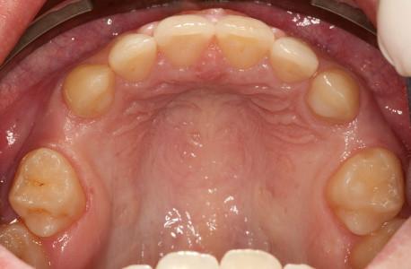
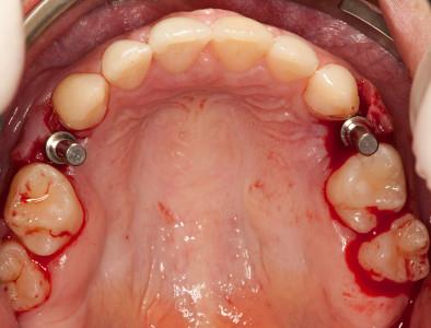
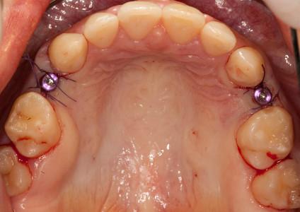
図1:口腔内写真:領域#14および#24 (#5 & 12 UNIV)の2カ所の間隙
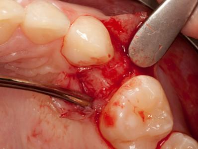
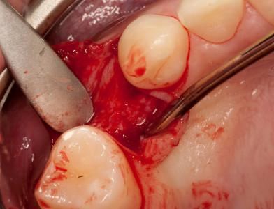
図2:領域#24 (#12 UNIV)における最小の粘膜骨膜弁の剥離
図3:領域#14 (#5 UNIV)における最小の粘膜骨膜弁の剥離
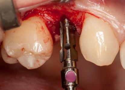
図4:インプラントの方向指示棒を用いてのポジション調整
図5:テーパードリル(ここではØ 3.5mm)を用いてのインプラント埋入部位のプレパレーション
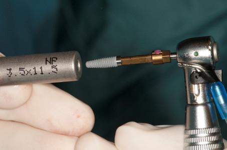
図6:インプラントをピックアップ
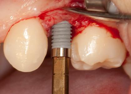
図7:インプラントを埋入(注:最大埋入トルクは45 Ncm)
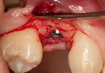
図8:やや歯槽骨縁下へのインプラント埋入(インプラント頚部が機械加工したカラーが保持されているため、骨吸収の場合に口腔衛生が容易)
図9:粘膜骨膜弁の再配置のために縫合
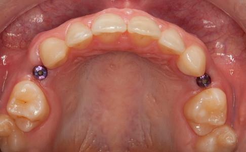
図10:インプラント埋入および抜歯から1週間後の口腔内
