-
0
Patient Assessment
- 0.1 Patient Demand
- 0.2 Anatomical location
-
0.3
Patient History
- 2.1 General patient history
- 2.2 Local history
-
0.4
Risk Assessment
- 3.1 Risk Assessment Overview
- 3.2 Age
- 3.3 Patient Compliance
- 3.4 Smoking
- 3.5 Drug Abuse
- 3.6 Recreational Drug and Alcohol Abuse
- 3.7 Condition of Natural Teeth
- 3.8 Parafunctions
- 3.9 Diabetes
- 3.10 Anticoagulants
- 3.11 Osteoporosis
- 3.12 Bisphosphonates
- 3.13 MRONJ
- 3.14 Steroids
- 3.15 Radiotherapy
- 3.16 Risk factors
-
1
Diagnostics
-
2
Treatment Options
-
2.1
Treatment planning
- 0.1 Non-implant based treatment options
- 0.2 Treatment planning conventional, model based, non-guided, semi-guided
- 0.3 Digital treatment planning
- 0.4 NobelClinician and digital workflow
- 0.5 Implant position considerations overview
- 0.6 Soft tissue condition and morphology
- 0.7 Site development, soft tissue management
- 0.8 Hard tissue and bone quality
- 0.9 Site development, hard tissue management
- 0.10 Time to function
- 0.11 Submerged vs non-submerged
- 0.12 Healed or fresh extraction socket
- 0.13 Screw-retained vs. cement-retained
- 0.14 Angulated Screw Channel system (ASC)
- 2.2 Treatment options esthetic zone
- 2.3 Treatment options posterior zone
- 2.4 Comprehensive treatment concepts
-
2.1
Treatment planning
-
3
Treatment Procedures
-
3.1
Treatment procedures general considerations
- 0.1 Anesthesia
- 0.2 peri-operative care
- 0.3 Flap- or flapless
- 0.4 Non-guided protocol
- 0.5 Semi-guided protocol
- 0.6 Guided protocol overview
- 0.7 Guided protocol NobelGuide
- 0.8 Parallel implant placement considerations
- 0.9 Tapered implant placement considerations
- 0.10 3D implant position
- 0.11 Implant insertion torque
- 0.12 Intra-operative complications
- 0.13 Impression procedures, digital impressions, intraoral scanning
- 3.2 Treatment procedures esthetic zone surgical
- 3.3 Treatment procedures esthetic zone prosthetic
- 3.4 Treatment procedures posterior zone surgical
- 3.5 Treatment procedures posterior zone prosthetic
-
3.1
Treatment procedures general considerations
-
4
Aftercare
Hard tissue augmentation (posterior zone)
Key points
- Vertical bone gain in posterior maxillae is mainly accomplished with a sinus augmentation procedure, while guided bone regeneration or a fixed crestal bone block serve the purpose in posterior mandibles.
- Sagittal bone gain in posterior jaw regions is mainly accomplished with a fixed bone block or with particulate bone attached to the buccal plate.
- Covering membranes are regularly used on grafting materials. Equally good results are seen with autografts, allografts, xenografts and alloplasts.
Posterior jaw regions
Loss of an upper single second bicuspid or molar may result in poor sagittal bone dimension, and also may induce an anterior/downward expansion of the sinus cavity revealing insufficient vertical bone dimension. The vertical bone height inferior to sinus with a minimum bucco-lingual dimension of 3.5-4.0 and 5.5 mm for a narrow/regular bicuspid or wide molar implant, respectively, will determine the type of implant procedure to be performed. No augmentation may require 8-10 mm available bone height for the single-tooth implant. Less vertical bone volume may call for an osteotome sinus floor augmentation technique via a crestal approach or a bone augmentation technique via a lateral sinus floor elevation approach.
Loss of a lower single second bicuspid or molar may reveal poor vertical and sagittal bone dimensions above the mental foramen and/or above the lower alveolar nerve. Short implants are preferably used whenever possible.
Reports on vertical and sagittal bone augmentation procedures at single tooth implants cover autografts, allografts, xenografts, and alloplasts, all with equally good results. No augmentation technique seems to be superior to others.
Surgical techniques
Vertical augmentation
The osteotome technique implies a drilling protocol to end a short distance from the sinus floor to be followed by careful hammering of osteotomes (various diameters ) to finally fracture the sinus floor upwards. The residual vertical bone height inferior to the sinus should preferably be >6mm. No bone augmentation material is required, but a vertical bone gain up to 4-5 mm may be accomplished and the implant is inserted during the same intervention.
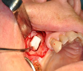
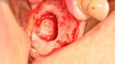
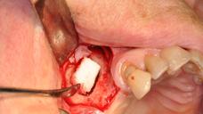
The lateral sinus floor elevation technique implies producing a buccal window into the sinus in the region of interest with conventional drills or with less invasive piezo-surgery. The aim is to lift an intact Schneiderian membrane and to either insert the implant immediately (residual bone inferior to sinus >3-4 mm) or, with less bone, just to fill the sinus with various bone material.
Figures 1 & 2: Lateral sinus floor elevation with bovine bone block/particles and covering collagen membrane
Vertical bone augmentation of the posterior mandible is mainly performed with guided bone regeneration using various membrane techniques or occasionally in some molar sites with crestal autogenous onlay bone blocks fixed with 1-2 mini-screws. Such procedures are challenging with risk of perforation of a thin and fragile mucosa, which will expose the graft.
Other techniques to improve the vertical dimension may be by means of vertical alveolar ridge distraction in either jaw, and in the mandible also by lateralization of the lower alveolar nerve, albeit these techniques are rarely performed in the single-tooth situation.
Sagittal augmentation
Lack of sagittal bone dimension will require buccal onlay augmentation, either with a mini-screw fixed autogenous bone block or with particulate bone substitute materials. Particulate bone may be kept in place using mini-screws as tent-poles, emerging 2-3 mm above the buccal bone plate. Membranes of various brands are regularly used with success to cover osteotomies and graft materials. Implants are generally placed according to a staged approach, i.e. 4-6 months post-grafting.
Digital Textbooks
Then, over time another phenomenon takes place. The maxillary sinus begins to enlarge and move downward in the direction of the residual ridge crest. This process of is known as pneumatization or "fourth expansion phenomenon" of the maxillary sinus. In the process, bone density in the affected areas also decreases rapidly along with bone quantity. The net result is formation of the least dense bony regions of the entire maxilla.


