-
0
Patient Assessment
- 0.1 Patient Demand
- 0.2 Anatomical location
-
0.3
Patient History
- 2.1 General patient history
- 2.2 Local history
-
0.4
Risk Assessment
- 3.1 Risk Assessment Overview
- 3.2 Age
- 3.3 Patient Compliance
- 3.4 Smoking
- 3.5 Drug Abuse
- 3.6 Recreational Drug and Alcohol Abuse
- 3.7 Condition of Natural Teeth
- 3.8 Parafunctions
- 3.9 Diabetes
- 3.10 Anticoagulants
- 3.11 Osteoporosis
- 3.12 Bisphosphonates
- 3.13 MRONJ
- 3.14 Steroids
- 3.15 Radiotherapy
- 3.16 Risk factors
-
1
Diagnostics
-
2
Treatment Options
-
2.1
Treatment planning
- 0.1 Non-implant based treatment options
- 0.2 Treatment planning conventional, model based, non-guided, semi-guided
- 0.3 Digital treatment planning
- 0.4 NobelClinician and digital workflow
- 0.5 Implant position considerations overview
- 0.6 Soft tissue condition and morphology
- 0.7 Site development, soft tissue management
- 0.8 Hard tissue and bone quality
- 0.9 Site development, hard tissue management
- 0.10 Time to function
- 0.11 Submerged vs non-submerged
- 0.12 Healed or fresh extraction socket
- 0.13 Screw-retained vs. cement-retained
- 0.14 Angulated Screw Channel system (ASC)
- 2.2 Treatment options esthetic zone
- 2.3 Treatment options posterior zone
- 2.4 Comprehensive treatment concepts
-
2.1
Treatment planning
-
3
Treatment Procedures
-
3.1
Treatment procedures general considerations
- 0.1 Anesthesia
- 0.2 peri-operative care
- 0.3 Flap- or flapless
- 0.4 Non-guided protocol
- 0.5 Semi-guided protocol
- 0.6 Guided protocol overview
- 0.7 Guided protocol NobelGuide
- 0.8 Parallel implant placement considerations
- 0.9 Tapered implant placement considerations
- 0.10 3D implant position
- 0.11 Implant insertion torque
- 0.12 Intra-operative complications
- 0.13 Impression procedures, digital impressions, intraoral scanning
- 3.2 Treatment procedures esthetic zone surgical
- 3.3 Treatment procedures esthetic zone prosthetic
- 3.4 Treatment procedures posterior zone surgical
- 3.5 Treatment procedures posterior zone prosthetic
-
3.1
Treatment procedures general considerations
-
4
Aftercare
Esthetic complications
Key points
- Esthetic complications are not uncommon.
- Complications can be a result of implant placement, healing or lack of patient compliance.
- Proper placement of the implant will minimize complications.
Esthetic complications and their management
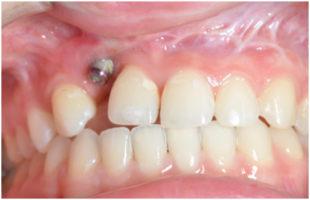
Esthetic complications in the anterior zone may result from a myriad of factors. The most obvious would be the poor placement of an implant. This could be a result of poor planning, lack of surgical guides, one not being familiar with the healing process, a lack of understanding of the implant to bone, adjacent teeth and / or soft tissue. The patients individual response to the procedure(s) may also play a role.
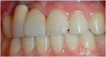
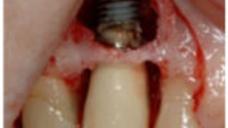
When implant placement is so compromised it may be necessary to remove the implant, graft, and start over. In clinical situations where the implant placement (for whatever the reason) is compromised there are workarounds with angulated abutments, ASC (Angulated Screw Channel) or custom abutments. While these make the fabrication of the restoration more challenging, the end result is often times acceptable to the patient. This can occur for the reasons mentioned above in addition to the fact that the osseous support only allowed for placement of the implant in that location.
Esthetic complications can also occur as a result of the implant being placed in the incorrect vertical positions (super crestal, crestal and sub crestal). Placing an implant in position that is too coronal many times compromises the final esthetic result by not providing sufficient restorative space of the components and materials necessary to create the final restoration.. This may also result in the actual implant platform being visible, a very difficult esthetic challenge. It is often times better error in placing the implant further apical.
Care must be given in preserving the inter-proximal (if present ) bone as this supports the soft tissue. Loss of this critical osseous support will result in the loss of the papilla, and potentially “black triangles”. Although this may be restored with pink materials it is difficult to do. Therefore a thorough understanding of the relationship of the implant to surrounding structures, teeth, adjacent implants, contact point, buccal plate, lingual plate or palate, and when to graft are all essential for an esthetic restoration.
There are also esthetic complications that occur after significant time as passed with the final restoration in place. These can result from residual cement, occlusion, screw loosening, poor fit of components, poor hygiene, lack of biocompatibility of materials, to list a few. The diagnosis of such a complication would then dictate the treatment.
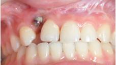
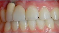
Figure 1: Compromised implant placement Figure 2: Even with angulated abutment, esthetics is compromised Figure 3: Bone loss due to incomplete cement removal
