-
0
Patient Assessment
- 0.1 Patient Demand
- 0.2 Anatomical location
-
0.3
Patient History
- 2.1 General patient history
- 2.2 Local history
-
0.4
Risk Assessment
- 3.1 Risk Assessment Overview
- 3.2 Age
- 3.3 Patient Compliance
- 3.4 Smoking
- 3.5 Drug Abuse
- 3.6 Recreational Drug and Alcohol Abuse
- 3.7 Condition of Natural Teeth
- 3.8 Parafunctions
- 3.9 Diabetes
- 3.10 Anticoagulants
- 3.11 Osteoporosis
- 3.12 Bisphosphonates
- 3.13 MRONJ
- 3.14 Steroids
- 3.15 Radiotherapy
- 3.16 Risk factors
-
1
Diagnostics
-
2
Treatment Options
-
2.1
Treatment planning
- 0.1 Non-implant based treatment options
- 0.2 Treatment planning conventional, model based, non-guided, semi-guided
- 0.3 Digital treatment planning
- 0.4 NobelClinician and digital workflow
- 0.5 Implant position considerations overview
- 0.6 Soft tissue condition and morphology
- 0.7 Site development, soft tissue management
- 0.8 Hard tissue and bone quality
- 0.9 Site development, hard tissue management
- 0.10 Time to function
- 0.11 Submerged vs non-submerged
- 0.12 Healed or fresh extraction socket
- 0.13 Screw-retained vs. cement-retained
- 0.14 Angulated Screw Channel system (ASC)
- 2.2 Treatment options esthetic zone
- 2.3 Treatment options posterior zone
- 2.4 Comprehensive treatment concepts
-
2.1
Treatment planning
-
3
Treatment Procedures
-
3.1
Treatment procedures general considerations
- 0.1 Anesthesia
- 0.2 peri-operative care
- 0.3 Flap- or flapless
- 0.4 Non-guided protocol
- 0.5 Semi-guided protocol
- 0.6 Guided protocol overview
- 0.7 Guided protocol NobelGuide
- 0.8 Parallel implant placement considerations
- 0.9 Tapered implant placement considerations
- 0.10 3D implant position
- 0.11 Implant insertion torque
- 0.12 Intra-operative complications
- 0.13 Impression procedures, digital impressions, intraoral scanning
- 3.2 Treatment procedures esthetic zone surgical
- 3.3 Treatment procedures esthetic zone prosthetic
- 3.4 Treatment procedures posterior zone surgical
- 3.5 Treatment procedures posterior zone prosthetic
-
3.1
Treatment procedures general considerations
-
4
Aftercare
Implant position (posterior zone)
Key points
- Mesio distal space (interdental space): 7mm (-> 1 implant), 12 mm (-> 2 implants).
- Bucco-palatal space: 6mm (regular implant: Ø 4mm).
- Distance from adjacent teeth: ≥1.5mm.
- Inter-implant space: 3mm.
- Inter-maxillary (occlusal) space: >10mm.
- Anatomical considerations: maxillary sinus, inferior alveolar nerve, mental nerve, lingual concavity (mandible).
- Alternatives: removable partial denture, resin-bonded fixed prosthesis, fixed partial denture.
Anatomical considerations
Prior to implant placement in the posterior site, a thorough radiographic examination by means of panoramic x-ray and/or dental CT or CBCT scan is recommended. This allows visualization of possible anatomic pitfalls, bone quantity and neuro-vascular structures. For panoramic x-rays, correction for the magnification factor should be applied. A safety margin of 2 mm between the apex of the implant and the nerve canal must be respected (Greenstein et al. 2008).
Clinical aspects
The clinical evaluation consists of measuring the mesio-distal space of the tooth gap (at least 7 mm for one implant or 12 mm for two implants), the bucco-palatal space (at least 6 mm to allow the insertion of a regular implant), the inter-occlusal space (at least 6 mm for fixed and 12 mm for removable dentures), bony defects after extraction which need augmentation, width of keratinized mucosa, gingival biotype (thin-scalloped or thick flat) and mouth-opening (>40mm).
Surgical protocol
Implant placement can be classified by timing: immediate, early and delayed. While immediate implant placement has several advantages for the patient, in particular reduction of treatment time, it is not always feasible and demands more surgical skills. Primary stability is easier accomplished in the molar site by placing the implant in the inter-radicular bony septum of a molar.
A knife-edged alveolar ridge (delayed implant placement) may necessarily need bone augmentation in advance or, in case of sufficient bone height, preparation of crestal bone to gain enough width for a regular implant dimension.
Implants are designed with and without machined collar. Studies, which compared bone loss between those two designs when placed at the crestal level, have shown that after an unloaded period, implants without machined collar had lost no bone. However, it is worthwhile mentioning, that the question of machined collar or not is still in debate, as crestal bone loss often occurs after loading and from a periodontal point of view, plaque may be easier removed from machined collars. Bone remodeling is observed around implants with machined collar, which are placed epicrestal. Bone-loss tends to be less when these implants are placed supracrestal.
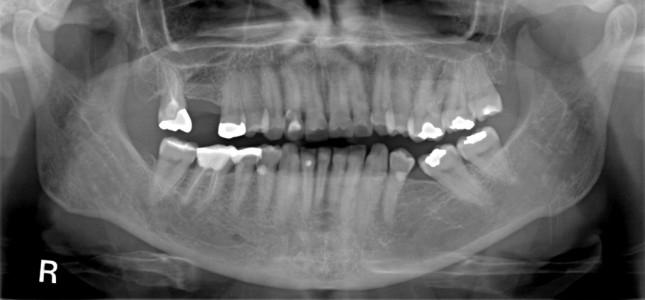
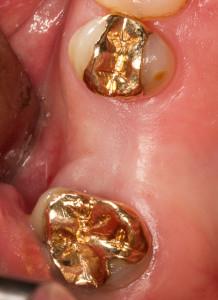
Figure 1: Missing tooth #16 (#3 UNIV)
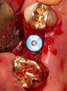
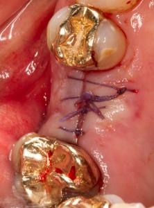
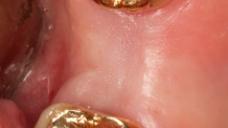
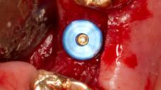
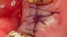
Figure 2: Occlusal view Figure 3: Wide platform placed in respect to adjacent teeth and buccal bone width Figure 4: Submerged healing
