-
0
Patient Assessment
- 0.1 Patient Demand
- 0.2 Anatomical location
-
0.3
Patient History
- 2.1 General patient history
- 2.2 Local history
-
0.4
Risk Assessment
- 3.1 Risk Assessment Overview
- 3.2 Age
- 3.3 Patient Compliance
- 3.4 Smoking
- 3.5 Drug Abuse
- 3.6 Recreational Drug and Alcohol Abuse
- 3.7 Condition of Natural Teeth
- 3.8 Parafunctions
- 3.9 Diabetes
- 3.10 Anticoagulants
- 3.11 Osteoporosis
- 3.12 Bisphosphonates
- 3.13 MRONJ
- 3.14 Steroids
- 3.15 Radiotherapy
- 3.16 Risk factors
-
1
Diagnostics
-
2
Treatment Options
-
2.1
Treatment planning
- 0.1 Non-implant based treatment options
- 0.2 Treatment planning conventional, model based, non-guided, semi-guided
- 0.3 Digital treatment planning
- 0.4 NobelClinician and digital workflow
- 0.5 Implant position considerations overview
- 0.6 Soft tissue condition and morphology
- 0.7 Site development, soft tissue management
- 0.8 Hard tissue and bone quality
- 0.9 Site development, hard tissue management
- 0.10 Time to function
- 0.11 Submerged vs non-submerged
- 0.12 Healed or fresh extraction socket
- 0.13 Screw-retained vs. cement-retained
- 0.14 Angulated Screw Channel system (ASC)
- 2.2 Treatment options esthetic zone
- 2.3 Treatment options posterior zone
- 2.4 Comprehensive treatment concepts
-
2.1
Treatment planning
-
3
Treatment Procedures
-
3.1
Treatment procedures general considerations
- 0.1 Anesthesia
- 0.2 peri-operative care
- 0.3 Flap- or flapless
- 0.4 Non-guided protocol
- 0.5 Semi-guided protocol
- 0.6 Guided protocol overview
- 0.7 Guided protocol NobelGuide
- 0.8 Parallel implant placement considerations
- 0.9 Tapered implant placement considerations
- 0.10 3D implant position
- 0.11 Implant insertion torque
- 0.12 Intra-operative complications
- 0.13 Impression procedures, digital impressions, intraoral scanning
- 3.2 Treatment procedures esthetic zone surgical
- 3.3 Treatment procedures esthetic zone prosthetic
- 3.4 Treatment procedures posterior zone surgical
- 3.5 Treatment procedures posterior zone prosthetic
-
3.1
Treatment procedures general considerations
-
4
Aftercare
Prosthodontic diagnostic tools overview
Key points
- Determination of available three-dimensional space for a single prosthetic reconstruction is an important item of pre-treatment diagnostics.
- Obtaining accurate diagnostic casts and an inter-occlusal/arch registration permits precise evaluation of existing morphological determinants and treatment possibilities.
Overview
Although rehabilitation with an implant-supported single-tooth restoration concerns only a small part of the total arch, it is important to diagnose available space and occlusion. Specific tools are available to enable determination of available three-dimensional space for a single prosthetic reconstruction and occlusion.
Methods are:
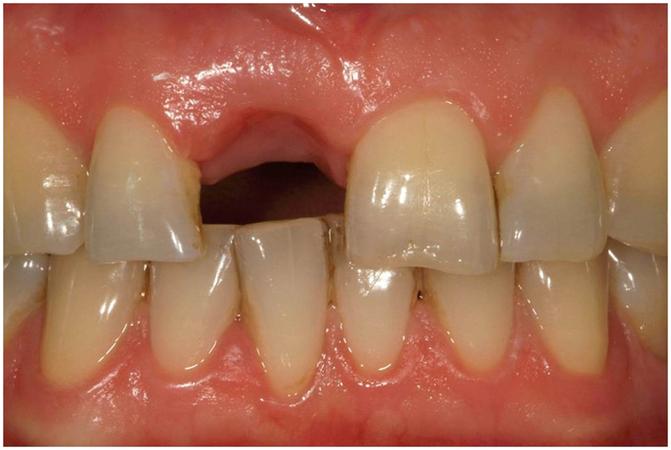
- determine the appropriate restorative space visually by comparing with the contralateral tooth. If the contralateral tooth has a normal width, it can often be used as a guide to determine sufficient space for the missing tooth;
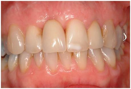
- inspect visually the dimensions of the temporary restoration to determine if it fills the extraction gap harmoniously;
- obtain computer models of both dental arches and their occlusal relationship by using an intraoral optical scanner; software programs can add teeth for diagnostic purposes;
- obtain gypsum casts and an inter-occlusal/arch registration; a conventional diagnostic wax-up can give insight in available dimensions;
- obtain gypsum casts and an inter-occlusal/arch registration; with optical scanning of the casts in the dental laboratory, computer models can be made and teeth can be added with a software program.
The above mentioned methods, together with cone beam computed tomography, gives sufficient insight in available space for the future restoration, available bone at the future implant site and relation to neighbouring anatomical structures. One must also keep in mind the cost-effectiveness for each diagnostic approach. Extensive and therefore expensive methods are not necessary in all cases.
Figure 1: Patient with a wish for implant-supported restorations in position 11 (#8 UNIV); method to determine the appropriate restorative space visually by comparing with the contralateral tooth.
Figure 2: Patient with a wish for implant-supported restorations 11 (#8 UNIV); method to inspect visually the dimensions of the temporary restoration to determine if it fills the extraction gap harmoniously.


Recent diagnostic tools in fixed prosthodontics