-
0
Patient Assessment
- 0.1 Patient Demand
- 0.2 Anatomical location
-
0.3
Patient History
- 2.1 General patient history
- 2.2 Local history
-
0.4
Risk Assessment
- 3.1 Risk Assessment Overview
- 3.2 Age
- 3.3 Patient Compliance
- 3.4 Smoking
- 3.5 Drug Abuse
- 3.6 Recreational Drug and Alcohol Abuse
- 3.7 Condition of Natural Teeth
- 3.8 Parafunctions
- 3.9 Diabetes
- 3.10 Anticoagulants
- 3.11 Osteoporosis
- 3.12 Bisphosphonates
- 3.13 MRONJ
- 3.14 Steroids
- 3.15 Radiotherapy
- 3.16 Risk factors
-
1
Diagnostics
-
2
Treatment Options
-
2.1
Treatment planning
- 0.1 Non-implant based treatment options
- 0.2 Treatment planning conventional, model based, non-guided, semi-guided
- 0.3 Digital treatment planning
- 0.4 NobelClinician and digital workflow
- 0.5 Implant position considerations overview
- 0.6 Soft tissue condition and morphology
- 0.7 Site development, soft tissue management
- 0.8 Hard tissue and bone quality
- 0.9 Site development, hard tissue management
- 0.10 Time to function
- 0.11 Submerged vs non-submerged
- 0.12 Healed or fresh extraction socket
- 0.13 Screw-retained vs. cement-retained
- 0.14 Angulated Screw Channel system (ASC)
- 2.2 Treatment options esthetic zone
- 2.3 Treatment options posterior zone
- 2.4 Comprehensive treatment concepts
-
2.1
Treatment planning
-
3
Treatment Procedures
-
3.1
Treatment procedures general considerations
- 0.1 Anesthesia
- 0.2 peri-operative care
- 0.3 Flap- or flapless
- 0.4 Non-guided protocol
- 0.5 Semi-guided protocol
- 0.6 Guided protocol overview
- 0.7 Guided protocol NobelGuide
- 0.8 Parallel implant placement considerations
- 0.9 Tapered implant placement considerations
- 0.10 3D implant position
- 0.11 Implant insertion torque
- 0.12 Intra-operative complications
- 0.13 Impression procedures, digital impressions, intraoral scanning
- 3.2 Treatment procedures esthetic zone surgical
- 3.3 Treatment procedures esthetic zone prosthetic
- 3.4 Treatment procedures posterior zone surgical
- 3.5 Treatment procedures posterior zone prosthetic
-
3.1
Treatment procedures general considerations
-
4
Aftercare
Dentogingival complex and biologic width
Key points
- The biological width is determined by the keratinized oral epithelium, the peri-implant junctional epithelium, and the sub-epithelial connective tissue.
- Gingiva and peri-implant mucosa differ in composition of cells, fibers, collagen, and vessels.
Differences between peri-implant mucosa and gingiva
The peri-implant mucosa consists of an externally located keratinized oral epithelium, which is connected to the peri-implant junctional epithelium facing the abutment. The latter extends approximately 2 mm apical to the coronal soft tissue margin, still being 1.0 –1.5 mm away from the peri-implant bone crest.
The biological width differs between implants and teeth, being >0.5 mm greater around implants (3.8 vs 3.2 mm), which is mainly due to the greater height of the interposed connective tissue.

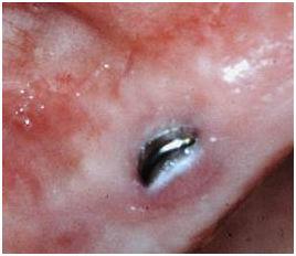
Figure 1: Accidental mucosal perforation of a submerged implant. This will frequently result in marginal bone resorption, assumingly aiming to adjust the biological width.
Compared to teeth, the supra-crestal connective tissue around implants contains fewer fibroblasts but more collagen fibers. In close vicinity to the implant/abutment surface, marginal bone attached collagen fibers run parallel to the implant/abutment surface. Fibers in gingiva around teeth are mainly circular and root cementum attached with a different orientation than around implants.
There are less vascular structures in the supra-crestal connective tissue near the implant compared to the same location around teeth.
The composition of cells, fibers, collagen, and vessels makes the peri-implant connective tissue more comparable to a scar tissue.
Substantial data support that implants of different materials (e.g titanium and zirconia) and of various roughness show a similar composition pattern, however with minor deviations.
