-
0
Patient Assessment
- 0.1 Patient demand
- 0.2 Overarching considerations
- 0.3 Local history
- 0.4 Anatomical location
- 0.5 General patient history
-
0.6
Risk assessment & special high risk categories
- 5.1 Risk assessment & special high risk categories
- 5.2 age
- 5.3 Compliance
- 5.4 Smoking
- 5.5 Drug abuse
- 5.6 Recreational drugs and alcohol abuse
- 5.7 Parafunctions
- 5.8 Diabetes
- 5.9 Osteoporosis
- 5.10 Coagulation disorders and anticoagulant therapy
- 5.11 Steroids
- 5.12 Bisphosphonates
- 5.13 BRONJ / ARONJ
- 5.14 Radiotherapy
- 5.15 Risk factors
-
1
Diagnostics
-
1.1
Clinical Assessment
- 0.1 Lip line
- 0.2 Mouth opening
- 0.3 Vertical dimension
- 0.4 Maxillo-mandibular relationship
- 0.5 TMD
- 0.6 Existing prosthesis
- 0.7 Muco-gingival junction
- 0.8 Hyposalivation and Xerostomia
- 1.2 Clinical findings
-
1.3
Clinical diagnostic assessments
- 2.1 Microbiology
- 2.2 Salivary output
-
1.4
Diagnostic imaging
- 3.1 Imaging overview
- 3.2 Intraoral radiographs
- 3.3 Panoramic
- 3.4 CBCT
- 3.5 CT
- 1.5 Diagnostic prosthodontic guides
-
1.1
Clinical Assessment
-
2
Treatment Options
- 2.1 Mucosally-supported
-
2.2
Implant-retained/supported, general
- 1.1 Prosthodontic options overview
- 1.2 Number of implants maxilla and mandible
- 1.3 Time to function
- 1.4 Submerged or non-submerged
- 1.5 Soft tissue management
- 1.6 Hard tissue management, mandible
- 1.7 Hard tissue management, maxilla
- 1.8 Need for grafting
- 1.9 Healed vs fresh extraction socket
- 1.10 Digital treatment planning protocols
- 2.3 Implant prosthetics - removable
-
2.4
Implant prosthetics - fixed
- 2.5 Comprehensive treatment concepts
-
3
Treatment Procedures
-
3.1
Surgical
-
3.2
Removable prosthetics
-
3.3
Fixed prosthetics
-
3.1
Surgical
- 4 Aftercare
Inflammation and infection
Key points
- Mucositis and Peri-implantitis treatments focus on elimination of pathogenic biofilm and removal of causal or supporting factors such as subgingival cement remnants, restoration cleansability, hygiene etc.
- Typical antimicrobials are 0.2 % Chlorhexidine, and adjunctive therapy options with antibiotics
- Consider increasing recall frequency and hygiene instructions to patient
- Adjunctive antimicrobial therapy options such as laser or ATP do not provide evidence-based or increased efficiency and efficacy
Peri-implant Inflammation, infection
Inflammation can be limited to the soft tissue around the implant (mucositis) or involve the underlying bone tissue as well (peri-implantitis). Infection can be caused by various risk factors, such as inadequate hygiene and patient compliance, inadequate restoration design to allow cleansing, aggressive oro-pharyngeal microflora, overloading of the implant, lack of fixed keratinized tissue, subingival cement remnants.
Plan regular hygiene recalls to check any inflammation signs. They can be subtle with light swelling, increased redness and discrete bleeding on probing (BOP). They can also be obvious with pain, distinct swelling and intense BOP, probing depth > 5mm, exsudation or even secretion of pus, clearly visible marginal bone loss. In extreme situations implant mobility may occur.
Therapy is depending on degree of inflammation. It is key to reduce/eliminate the microbial load, as well as other causes for inflammation (e.g. subgingival cement excess)
Mucositis
Treatment consists of cleaning of prosthetic components (including bar, attachments), supra and- subgingival implant surfaces, plaque and calculus removal, eventually removal of inflamed tissues.
Rinse and disinfect periimplant pocket with antimicrobial solution, such as 0.2 % chlorhexidine or 2 % hydrogen-peroxide
Check design of restoration for cleansablity, occlusion and articulation. If needed eliminate premature contacts and implant overload. If necessary, consider design adaptation or even remake of prosthesis with more adequate cleansability
Hygiene re-instruction of patient is a must
Prescribe 0.2 % Chlorhexidine oral rinses, 3 times daily
Recall and reevaluate after 2 weeks. Consider increasing recall frequency and hygiene instructions.
Even if hard data on use of local antibiotics are lacking, in case of recurrence of infection consider local application of concentrated antibiotic gels (Metronidazole, Minocycline, Tetracycline).
If arguments for candida or other yeast infection arise prescribe the appropriate anti yeast medication like miconazole.
Peri-implantitis
Based on standardized intra-oral or panoramic radiographs revealing a loss of marginal bone over time.
Treatment consists of :
Removal of restoration/overdenture, hygiene and disinfection measures, evaluation/adaptation of prosthetic restoration evaluation as described above
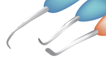
Removal of microbial biofilms, cleaning of implant surfaces with suitable plastic or titanium scalers and curettes and removal of infected soft tissue, if necessary implantoplasty with suitable diamond burs and implantoplasty brushes. Irrigation and disinfection of periimplant site with 0.2 % Chlorhexidine.
If needed peri-implant soft tissue management and creation of sufficient keratinized gingiva may be an option
If indicated, resective and regenerative treatment of bony defects
In some cases explantation and revision surgery need to be considered.
Antibiosis
Local application of concentrated antibiotic gels (Metronidazole, Tetracycline) has been tried out with variable success.
Gramnegative anaerobe microorganisms are frequently involved. For periimplantitis the effects of a systemic antibiotics, as adjunct to mechanical/surgical treatment remain unknown. Microbial testing can help selecting a proper antibiotic by detecting resistant species.
Typical antibiotics used are Metronidazole (3x 400 mg/d), Clindamycine (4 x 300 mg/d), and Amoxicilline (3 x 500 mg/d) (in case of patient intolerance against Penicillin, use Ciprofloxacine). Combination of Amoxicilline (3 x 500/d) and Metronidazole (3 x 400/d) is also effective against A.actinomycetemcomitans (or Ciprofloxacine 2 x 250 mg/d and Metronidazole 2 x 500mg/d)
Other adjunctive antimicrobial treatment measures
Use of abrasive powder airpolishing devices should be done, if at all, with great prudence due to risk of leaving powder remnants in the tissues or provoking an emphysema.
Effectiveness and efficiency of Antimicrobial Photodynamic Therapy remains controversial.
Co2 and Er:YAG Laser: Lasers seem to have an antimicrobial effect in vitro. However there is no clinical evidence Laser decontamination would be effective or even more effective as as other protocols used.

