-
0
Patient Assessment
- 0.1 Patient demand
- 0.2 Overarching considerations
- 0.3 Local history
- 0.4 Anatomical location
- 0.5 General patient history
-
0.6
Risk assessment & special high risk categories
- 5.1 Risk assessment & special high risk categories
- 5.2 age
- 5.3 Compliance
- 5.4 Smoking
- 5.5 Drug abuse
- 5.6 Recreational drugs and alcohol abuse
- 5.7 Parafunctions
- 5.8 Diabetes
- 5.9 Osteoporosis
- 5.10 Coagulation disorders and anticoagulant therapy
- 5.11 Steroids
- 5.12 Bisphosphonates
- 5.13 BRONJ / ARONJ
- 5.14 Radiotherapy
- 5.15 Risk factors
-
1
Diagnostics
-
1.1
Clinical Assessment
- 0.1 Lip line
- 0.2 Mouth opening
- 0.3 Vertical dimension
- 0.4 Maxillo-mandibular relationship
- 0.5 TMD
- 0.6 Existing prosthesis
- 0.7 Muco-gingival junction
- 0.8 Hyposalivation and Xerostomia
- 1.2 Clinical findings
-
1.3
Clinical diagnostic assessments
- 2.1 Microbiology
- 2.2 Salivary output
-
1.4
Diagnostic imaging
- 3.1 Imaging overview
- 3.2 Intraoral radiographs
- 3.3 Panoramic
- 3.4 CBCT
- 3.5 CT
- 1.5 Diagnostic prosthodontic guides
-
1.1
Clinical Assessment
-
2
Treatment Options
- 2.1 Mucosally-supported
-
2.2
Implant-retained/supported, general
- 1.1 Prosthodontic options overview
- 1.2 Number of implants maxilla and mandible
- 1.3 Time to function
- 1.4 Submerged or non-submerged
- 1.5 Soft tissue management
- 1.6 Hard tissue management, mandible
- 1.7 Hard tissue management, maxilla
- 1.8 Need for grafting
- 1.9 Healed vs fresh extraction socket
- 1.10 Digital treatment planning protocols
- 2.3 Implant prosthetics - removable
-
2.4
Implant prosthetics - fixed
- 2.5 Comprehensive treatment concepts
-
3
Treatment Procedures
-
3.1
Surgical
-
3.2
Removable prosthetics
-
3.3
Fixed prosthetics
-
3.1
Surgical
- 4 Aftercare
全口义齿印模
Key points
- 印模的目的是记录并传输软组织形态的口腔情况
- 需要获得义齿基托延伸相对于患者机能性运动的精确记录
- 通过口腔扫描技术获得的新型数字化印模可能会使传统印模技术过时
- 数字化印模的优势表现在数字化工作流程方面,可以为患者和临床医师提供治疗便利
软组织表面记录
针对全口义齿制作的印模记录可用的义齿支撑组织表面。 需要获得基于支撑组织负荷支撑能力的理想力量分布模型,同时避免义齿基托的口面肌肉移动。
传统印模
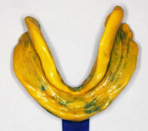
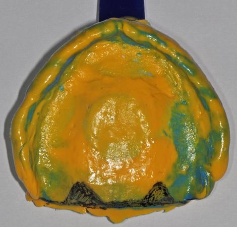
标准全口义齿印模技术涉及专门设计的刚性定制托盘和弹性印模材料,顺序如下:
1. 刚性印模托盘在具有相应蜡塑浮雕的石膏模型上制成。在理想情况下,蜡塑浮雕产生的空间可以使印模材料厚度均匀。
2. 可以使用建模复合物或弹性材料塑造印模托盘边缘,以获取患者口腔肌肉的功能性运动。 下颌舌部延伸需要特别注意,因为舌头肌肉活动强烈。 印模托盘随后将会加载材料,并按照制造商建议的时间段插入。
3. 设置弹性材料之后,取下印模,用牙科石膏浇注印模。
数字化印模
口腔扫描仪也可以为技师提供虚拟主模型,而通过打磨、打印或立体平版印刷技术,虚拟主模型可以转换成模拟主模型。 此类扫描仪的逐渐采用最终将会取代传统印模技术。
