-
0
Patient Assessment
- 0.1 Patient demand
- 0.2 Overarching considerations
- 0.3 Local history
- 0.4 Anatomical location
- 0.5 General patient history
-
0.6
Risk assessment & special high risk categories
- 5.1 Risk assessment & special high risk categories
- 5.2 age
- 5.3 Compliance
- 5.4 Smoking
- 5.5 Drug abuse
- 5.6 Recreational drugs and alcohol abuse
- 5.7 Parafunctions
- 5.8 Diabetes
- 5.9 Osteoporosis
- 5.10 Coagulation disorders and anticoagulant therapy
- 5.11 Steroids
- 5.12 Bisphosphonates
- 5.13 BRONJ / ARONJ
- 5.14 Radiotherapy
- 5.15 Risk factors
-
1
Diagnostics
-
1.1
Clinical Assessment
- 0.1 Lip line
- 0.2 Mouth opening
- 0.3 Vertical dimension
- 0.4 Maxillo-mandibular relationship
- 0.5 TMD
- 0.6 Existing prosthesis
- 0.7 Muco-gingival junction
- 0.8 Hyposalivation and Xerostomia
- 1.2 Clinical findings
-
1.3
Clinical diagnostic assessments
- 2.1 Microbiology
- 2.2 Salivary output
-
1.4
Diagnostic imaging
- 3.1 Imaging overview
- 3.2 Intraoral radiographs
- 3.3 Panoramic
- 3.4 CBCT
- 3.5 CT
- 1.5 Diagnostic prosthodontic guides
-
1.1
Clinical Assessment
-
2
Treatment Options
- 2.1 Mucosally-supported
-
2.2
Implant-retained/supported, general
- 1.1 Prosthodontic options overview
- 1.2 Number of implants maxilla and mandible
- 1.3 Time to function
- 1.4 Submerged or non-submerged
- 1.5 Soft tissue management
- 1.6 Hard tissue management, mandible
- 1.7 Hard tissue management, maxilla
- 1.8 Need for grafting
- 1.9 Healed vs fresh extraction socket
- 1.10 Digital treatment planning protocols
- 2.3 Implant prosthetics - removable
-
2.4
Implant prosthetics - fixed
- 2.5 Comprehensive treatment concepts
-
3
Treatment Procedures
-
3.1
Surgical
-
3.2
Removable prosthetics
-
3.3
Fixed prosthetics
-
3.1
Surgical
- 4 Aftercare
Complete denture impressions
Key points
- The objective of the impression is to record and transfer the intraoral situation of the soft tissue morphology
- Accurate recording of denture base extensions relative to functional movements of the patient need to be captured
- New digital impressions taken via intra-oral scanning techniques may make traditional impression-taking techniques obsolete
- Digital impressions offer advantages in terms of the digital workflow and provide treatment convenience for both patient and clinician
Soft tissue surface recording
Impression taking for a complete denture records available denture-bearing tissue surfaces. An ideal force distribution pattern based upon the load bearing capacity of supporting tissues is desirable while avoiding dislodging by the oro-facial musculature of the denture bases.
Conventional impressions
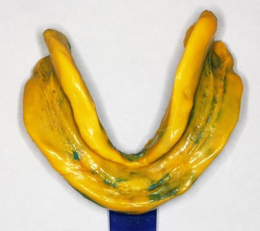
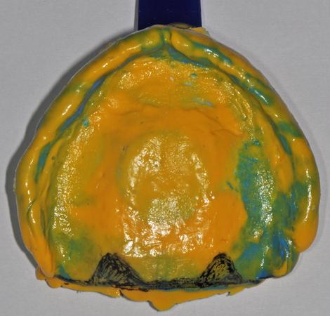
A standard complete denture impression technique involves specially designed rigid custom trays and resilient impression materials in the following sequence:
- Rigid impression trays made on stone casts with appropriate wax relief. Ideally, wax relief creates space for an even thickness of impression material.
- The impression tray may be border-molded using modeling compound or elastomeric materials in order to capture functional movements of the patient's intra-oral musculature. The mandibular lingual extension deserves particular attention due to the strong activity of the tongue muscles. The impression tray is then loaded with material and inserted for the time period recommended by the manufacturer.
- Once the resilient material has set, the impression is removed and the impressions are poured in dental stone.
Digital impressions
Intraoral scanners are also used to provide technicians with a virtual master cast that is converted into an analog cast via milling, printing or stereo-lithographic technique. Evolving use of such scanners may eventually eclipse traditional impression making techniques.
