-
0
Patient Assessment
- 0.1 Patient demand
- 0.2 Overarching considerations
- 0.3 Local history
- 0.4 Anatomical location
- 0.5 General patient history
-
0.6
Risk assessment & special high risk categories
- 5.1 Risk assessment & special high risk categories
- 5.2 age
- 5.3 Compliance
- 5.4 Smoking
- 5.5 Drug abuse
- 5.6 Recreational drugs and alcohol abuse
- 5.7 Parafunctions
- 5.8 Diabetes
- 5.9 Osteoporosis
- 5.10 Coagulation disorders and anticoagulant therapy
- 5.11 Steroids
- 5.12 Bisphosphonates
- 5.13 BRONJ / ARONJ
- 5.14 Radiotherapy
- 5.15 Risk factors
-
1
Diagnostics
-
1.1
Clinical Assessment
- 0.1 Lip line
- 0.2 Mouth opening
- 0.3 Vertical dimension
- 0.4 Maxillo-mandibular relationship
- 0.5 TMD
- 0.6 Existing prosthesis
- 0.7 Muco-gingival junction
- 0.8 Hyposalivation and Xerostomia
- 1.2 Clinical findings
-
1.3
Clinical diagnostic assessments
- 2.1 Microbiology
- 2.2 Salivary output
-
1.4
Diagnostic imaging
- 3.1 Imaging overview
- 3.2 Intraoral radiographs
- 3.3 Panoramic
- 3.4 CBCT
- 3.5 CT
- 1.5 Diagnostic prosthodontic guides
-
1.1
Clinical Assessment
-
2
Treatment Options
- 2.1 Mucosally-supported
-
2.2
Implant-retained/supported, general
- 1.1 Prosthodontic options overview
- 1.2 Number of implants maxilla and mandible
- 1.3 Time to function
- 1.4 Submerged or non-submerged
- 1.5 Soft tissue management
- 1.6 Hard tissue management, mandible
- 1.7 Hard tissue management, maxilla
- 1.8 Need for grafting
- 1.9 Healed vs fresh extraction socket
- 1.10 Digital treatment planning protocols
- 2.3 Implant prosthetics - removable
-
2.4
Implant prosthetics - fixed
- 2.5 Comprehensive treatment concepts
-
3
Treatment Procedures
-
3.1
Surgical
-
3.2
Removable prosthetics
-
3.3
Fixed prosthetics
-
3.1
Surgical
- 4 Aftercare
CBCT
Key points
- 与螺旋 CT 扫描相比,CBCT 的辐射剂量更低
- 辐射剂量取决于 mA 和辐射接触时间,而不是体素大小
- 经证明,尺寸精度较高
- 根据临床适应证,不同的硬件提供的视野也不同(如 4 x 4 到 15 x 20 厘米)
- 可获得 2D 图像和 3D 图像
- Hounsfield 单位测量值没有螺旋 CT 可靠
- 在图像中整合规划或现有的修复设备对于治疗方案设计并无价值
CBCT 注意事项
- 辐射源在其源头准直,然后发散为扇形以到达探测器。这会产生锥形束。
- 像素(图像 + 元素)是 2-D 图像最小的单个组成部分,而体素是 3-D 环境中最小的要素。
- 较大的视野 (FOV) 有助于观察颌骨及其关系。
- 小 FOV 则提供了骨小梁和皮质的详细图像。
- 图像内特定组织的灰度值各不相同,且不像在螺旋 CT 中那样可靠。
- CBCT 可以检测通过全景/口腔内 X 光片无法看到的临床相关结构,如切牙根管、舌骨凹部及缺失的骨皮质
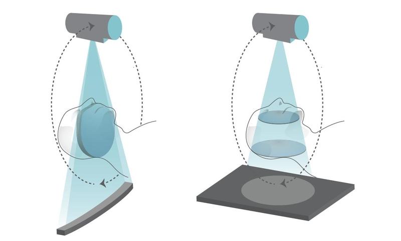
- CBCT 2-D 图像,显示切牙根管的近中延伸部
- CBCT 对术前规划以及钻针引导器和/或修复体的 CAD-CAM 制造是必不可少的
- 除灰度值外,CBCT 提供的信息准确度与螺旋 CT 相同甚至更高,则辐射接触量小,成本更低


