-
0
Patient Assessment
- 0.1 Patient Demand
- 0.2 Anatomical location
-
0.3
Patient History
- 2.1 General patient history
- 2.2 Local history
-
0.4
Risk Assessment
- 3.1 Risk Assessment Overview
- 3.2 Age
- 3.3 Patient Compliance
- 3.4 Smoking
- 3.5 Drug Abuse
- 3.6 Recreational Drug and Alcohol Abuse
- 3.7 Condition of Natural Teeth
- 3.8 Parafunctions
- 3.9 Diabetes
- 3.10 Anticoagulants
- 3.11 Osteoporosis
- 3.12 Bisphosphonates
- 3.13 MRONJ
- 3.14 Steroids
- 3.15 Radiotherapy
- 3.16 Risk factors
-
1
Diagnostics
-
2
Treatment Options
-
2.1
Treatment planning
- 0.1 Non-implant based treatment options
- 0.2 Treatment planning conventional, model based, non-guided, semi-guided
- 0.3 Digital treatment planning
- 0.4 NobelClinician and digital workflow
- 0.5 Implant position considerations overview
- 0.6 Soft tissue condition and morphology
- 0.7 Site development, soft tissue management
- 0.8 Hard tissue and bone quality
- 0.9 Site development, hard tissue management
- 0.10 Time to function
- 0.11 Submerged vs non-submerged
- 0.12 Healed or fresh extraction socket
- 0.13 Screw-retained vs. cement-retained
- 0.14 Angulated Screw Channel system (ASC)
- 2.2 Treatment options esthetic zone
- 2.3 Treatment options posterior zone
- 2.4 Comprehensive treatment concepts
-
2.1
Treatment planning
-
3
Treatment Procedures
-
3.1
Treatment procedures general considerations
- 0.1 Anesthesia
- 0.2 peri-operative care
- 0.3 Flap- or flapless
- 0.4 Non-guided protocol
- 0.5 Semi-guided protocol
- 0.6 Guided protocol overview
- 0.7 Guided protocol NobelGuide
- 0.8 Parallel implant placement considerations
- 0.9 Tapered implant placement considerations
- 0.10 3D implant position
- 0.11 Implant insertion torque
- 0.12 Intra-operative complications
- 0.13 Impression procedures, digital impressions, intraoral scanning
- 3.2 Treatment procedures esthetic zone surgical
- 3.3 Treatment procedures esthetic zone prosthetic
- 3.4 Treatment procedures posterior zone surgical
- 3.5 Treatment procedures posterior zone prosthetic
-
3.1
Treatment procedures general considerations
-
4
Aftercare
口内法X線撮影
Key points
- 口内法は、近遠心(水平)方向および頂部-根尖(垂直)方向の測定値に加え、骨の構造や密度に関しても有益な情報が得られます。
- もう1つの利点は、デジタル処理により画像を強調できることです。
- 他の方法を行う前に必ず平行法を試みます。
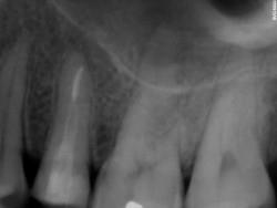
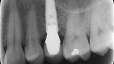
歯科領域では、口腔内X線撮影は依然として最も重要な撮影方法の1つです。口内法は、歯の構造や歯と顎骨の疾患を高い空間分解能で撮影します。口内法X線撮影は、注意深いキャリブレーションと検出器の入念なポジショニングにより、インプラント治療の計画作成に不可欠の診断情報を提供します。近遠心(水平)方向および頂部-根尖(垂直)方向の測定値に加え、骨の構造や密度に関しても有益な情報が得られます。
口内法の全顎撮影は、初診またはフォローアップで行われることが多く、14~21枚のX線写真を撮影します。また、根尖部の口内法X線写真は、以後のX線写真との比較により、経時的変化をモニタリングするためのベースライン基準を提供します。インプラント周囲の辺縁骨レベルは、インプラント周囲の骨の健康状態をモニタリングするための最も重要な方法の1つであり、インプラント成功の指標としても用いられています。
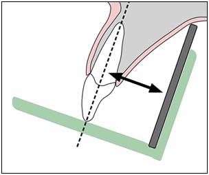
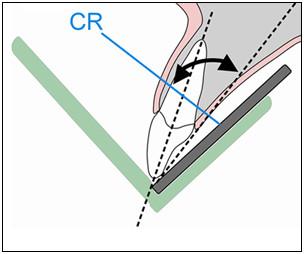
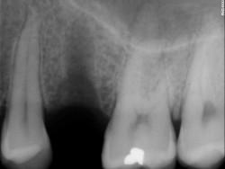
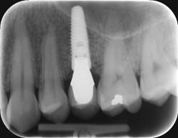
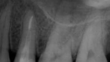
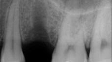
根尖部の口内法撮影では、平行法と二等分法の2種類の方法が用いられています(図1および図2)。平行法は、歯とセンサー面が平行になるように配置します。平行法は画像の歪みが少なく、患者の余剰な放射線被曝を低減します。
二等分法は、レセプターをできるだけ歯に近接させて配置し、歯軸とレセプター面によって形成される角度を二等分する線と垂直にX線の中心線を入射させます。この方法は、平行法ではアクセスが難しく、歯とフィルムの間に15度以上の角度を形成できない部位に用いられます。
他の方法を行う前に、必ず平行法を試みます。歯槽頂が著しく萎縮している場合は、歪みのリスクを低減するため、小さいサイズの検出器を使用します。
技術の進歩とともに、デジタル検出器は高画質を維持しつつ、放射線量を低減するための重要な要因となることが認識され、徐々に人気を博しています。もう1つの利点は、デジタル処理により画像を強調できることです。

About X-ray dental intraoral
Hello!If you can help me with something. Should the room where X-ray dental intraoral is located be isolated with Pb to work with it, or it can be used in normal dental ordinance without isolation?
Hello!
If you can help me with something. Should the room where X-ray dental intraoral is located be isolated with Pb to work with it, or it can be used in normal dental ordinance without isolation?