-
0
Patient Assessment
- 0.1 Patient Demand
- 0.2 Anatomical location
-
0.3
Patient History
- 2.1 General patient history
- 2.2 Local history
-
0.4
Risk Assessment
- 3.1 Risk Assessment Overview
- 3.2 Age
- 3.3 Patient Compliance
- 3.4 Smoking
- 3.5 Drug Abuse
- 3.6 Recreational Drug and Alcohol Abuse
- 3.7 Condition of Natural Teeth
- 3.8 Parafunctions
- 3.9 Diabetes
- 3.10 Anticoagulants
- 3.11 Osteoporosis
- 3.12 Bisphosphonates
- 3.13 MRONJ
- 3.14 Steroids
- 3.15 Radiotherapy
- 3.16 Risk factors
-
1
Diagnostics
-
2
Treatment Options
-
2.1
Treatment planning
- 0.1 Non-implant based treatment options
- 0.2 Treatment planning conventional, model based, non-guided, semi-guided
- 0.3 Digital treatment planning
- 0.4 NobelClinician and digital workflow
- 0.5 Implant position considerations overview
- 0.6 Soft tissue condition and morphology
- 0.7 Site development, soft tissue management
- 0.8 Hard tissue and bone quality
- 0.9 Site development, hard tissue management
- 0.10 Time to function
- 0.11 Submerged vs non-submerged
- 0.12 Healed or fresh extraction socket
- 0.13 Screw-retained vs. cement-retained
- 0.14 Angulated Screw Channel system (ASC)
- 2.2 Treatment options esthetic zone
- 2.3 Treatment options posterior zone
- 2.4 Comprehensive treatment concepts
-
2.1
Treatment planning
-
3
Treatment Procedures
-
3.1
Treatment procedures general considerations
- 0.1 Anesthesia
- 0.2 peri-operative care
- 0.3 Flap- or flapless
- 0.4 Non-guided protocol
- 0.5 Semi-guided protocol
- 0.6 Guided protocol overview
- 0.7 Guided protocol NobelGuide
- 0.8 Parallel implant placement considerations
- 0.9 Tapered implant placement considerations
- 0.10 3D implant position
- 0.11 Implant insertion torque
- 0.12 Intra-operative complications
- 0.13 Impression procedures, digital impressions, intraoral scanning
- 3.2 Treatment procedures esthetic zone surgical
- 3.3 Treatment procedures esthetic zone prosthetic
- 3.4 Treatment procedures posterior zone surgical
- 3.5 Treatment procedures posterior zone prosthetic
-
3.1
Treatment procedures general considerations
-
4
Aftercare
治癒済みまたは新たな抜歯窩
Key points
- 治癒済み抜歯窩と新たな抜歯窩とを比較した場合、埋入したインプラントの短期的残存率は同等のようです。
- 除去した歯根とそれよりも細い即時埋入インプラントとの間のサイズの不一致に関しては、骨領域を保護するため、しばしば骨片で間隙を埋める必要があります。
- この問題に関する長期的データは限られています
はじめに
単独歯インプラントは、しばしば切歯、犬歯、小臼歯の抜歯後に即時埋入されます。こうした手法は、多くの場合に即時負荷または早期負荷とともに、抜歯窩の壁を保護した慎重な抜歯および感染対策により実現します。2~3の歯根の臼歯を除去すると大きな骨欠損が残ることから、段階的アプローチが広く利用されています。これは、インプラントの初期固定を得る難しさと同時に、埋入されたインプラントを覆う軟組織量の不足によるものです。短期的な追跡調査においては、インプラントを新たな抜歯窩に埋入した場合と治癒済み抜歯窩に埋入した場合との間に差は見られませんでした。最近の文献では、インプラントの即時埋入を即時負荷と組み合わせた場合についても、インプラントの残存率を低下させないことが示されています。
新たな抜歯窩へのインプラントの即時埋入は、代用骨またはコラーゲン膜を用いた誘導骨再生が歯槽堤の高さおよび幅の維持を向上させる可能性はあるものの、歯槽頂の吸収を防止することはできません。しかし、インプラントの即時埋入の利点は、インプラント周囲軟組織を一定程度維持することにあります。新たな抜歯窩と治癒済み抜歯窩に埋入されたインプラントを比較した場合、2年後のインプラント周囲の骨吸収率は同等です。
治療済み抜歯窩へのインプラント
長所
- 感染可能性の解決
- 骨欠損の治癒
- 抜歯窩の軟組織治癒による軟組織量の増加
短所
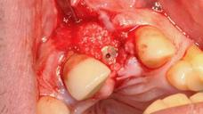
- 歯槽堤吸収が継続することによる骨量減少の進行。異種骨の小片等で歯槽を充填することにより、部分的に防止できる可能性があります。
- 治療期間の長期化
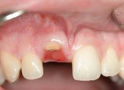
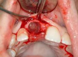
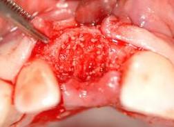
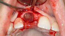
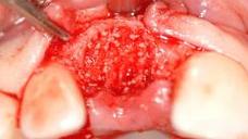
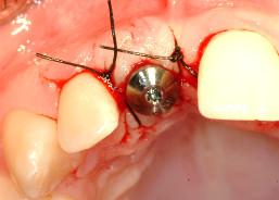
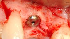
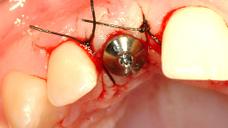
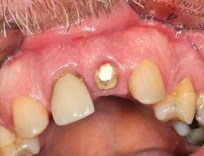
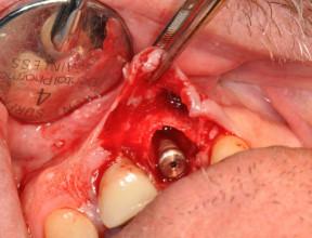
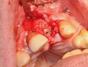
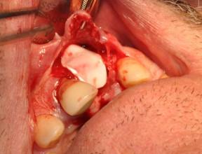
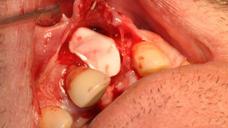
図3:異種骨を充填し、コラーゲン膜で覆った
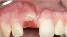
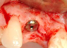
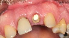
歯槽図6:インプラント埋入の2カ月後に装着した
ヒーリング・アバットメント新たな抜歯窩へのインプラント
長所
- 治療期間全体の短縮
- 患者の苦痛低減
短所
- 初期固定が低下する可能性
- 頬側/舌側部位のインプラント上の角化粘膜の量が減少する可能性
Digital Textbooks
Over the past 20 years there have been significant changes from the original Brånemark implant treatment protocols. Perhaps foremost has been immediate implant placement at the time of extraction which has become a viable treatment method. In addition, barrier membranes and grafting materials have been successfully used in conjunction with the immediate placement to augment extraction sockets and bony defects adjacent to immediately-placed implants.
Questions
ログインまたはご登録してコメントを投稿してください。
質問する
ログインまたは、無料でご登録して続行してください
You have reached the limit of content accessible without log in or this content requires log in. Log in or sign up now to get unlimited access to all FOR online resources.
FORウェブサイトにご登録していただきますと、すべてのオンライン・リソースに無制限にアクセスできます。FORウェブサイトへのご登録は無料となっております。
