-
0
Patient Assessment
- 0.1 Patient Demand
- 0.2 Anatomical location
-
0.3
Patient History
- 2.1 General patient history
- 2.2 Local history
-
0.4
Risk Assessment
- 3.1 Risk Assessment Overview
- 3.2 Age
- 3.3 Patient Compliance
- 3.4 Smoking
- 3.5 Drug Abuse
- 3.6 Recreational Drug and Alcohol Abuse
- 3.7 Condition of Natural Teeth
- 3.8 Parafunctions
- 3.9 Diabetes
- 3.10 Anticoagulants
- 3.11 Osteoporosis
- 3.12 Bisphosphonates
- 3.13 MRONJ
- 3.14 Steroids
- 3.15 Radiotherapy
- 3.16 Risk factors
-
1
Diagnostics
-
2
Treatment Options
-
2.1
Treatment planning
- 0.1 Non-implant based treatment options
- 0.2 Treatment planning conventional, model based, non-guided, semi-guided
- 0.3 Digital treatment planning
- 0.4 NobelClinician and digital workflow
- 0.5 Implant position considerations overview
- 0.6 Soft tissue condition and morphology
- 0.7 Site development, soft tissue management
- 0.8 Hard tissue and bone quality
- 0.9 Site development, hard tissue management
- 0.10 Time to function
- 0.11 Submerged vs non-submerged
- 0.12 Healed or fresh extraction socket
- 0.13 Screw-retained vs. cement-retained
- 0.14 Angulated Screw Channel system (ASC)
- 2.2 Treatment options esthetic zone
- 2.3 Treatment options posterior zone
- 2.4 Comprehensive treatment concepts
-
2.1
Treatment planning
-
3
Treatment Procedures
-
3.1
Treatment procedures general considerations
- 0.1 Anesthesia
- 0.2 peri-operative care
- 0.3 Flap- or flapless
- 0.4 Non-guided protocol
- 0.5 Semi-guided protocol
- 0.6 Guided protocol overview
- 0.7 Guided protocol NobelGuide
- 0.8 Parallel implant placement considerations
- 0.9 Tapered implant placement considerations
- 0.10 3D implant position
- 0.11 Implant insertion torque
- 0.12 Intra-operative complications
- 0.13 Impression procedures, digital impressions, intraoral scanning
- 3.2 Treatment procedures esthetic zone surgical
- 3.3 Treatment procedures esthetic zone prosthetic
- 3.4 Treatment procedures posterior zone surgical
- 3.5 Treatment procedures posterior zone prosthetic
-
3.1
Treatment procedures general considerations
-
4
Aftercare
Hard tissue augmentation (esthetic zone)
Key points
- Vertical bone gain in anterior maxillae for the single-tooth situation is mainly accomplished with guided bone regeneration techniques or with an L-shaped fixed bone block. Corresponding procedures in mandibles are rarely performed and merely possible to accomplish in the canine region.
- Sagittal bone gain in anterior jaw regions is mainly accomplished with a fixed bone block or with particulate bone, attached to the buccal plate.
- Covering membranes are regularly used on grafting materials. Equally good results are seen with autografts, allografts, xenografts and alloplasts.
Anterior jaw regions
Loss of an upper incisor or canine may frequently reveal insufficient vertical bone dimension, and often a poor sagittal bone dimension. Extraction of the central incisor may bring about expansion of the incisive foramen, leaving insufficient residual bone volume to accommodate an implant. Vertical bone height inferior to the nasal cavity with a minimum bucco-lingual dimension of 3.5-4.0 mm for a narrow/regular incisor or canine implant will determine the type of implant procedure to be performed. A straightforward operation (no augmentation) may require 8-10 mm available bone height for the single-tooth implant. Less vertical bone volume will call for an augmentation procedure of the sagittal and/or vertical bone dimension.
The handling of the lower canine region is comparable to the upper one, although loss of a lower single incisor will often lead to a compromised situation with poor bone volume in all three dimensions, especially so with a reduced mesio-distal space. Always consider orthodontic treatment or a resin-bonded fixed partial denture as alternative concepts.
Reports on vertical and sagittal bone augmentation procedures at single tooth implants cover autografts, allografts, xenografts, and alloplasts, all with equally good results. No augmentation technique seems to be superior to others.
Surgical techniques
Vertical augmentation
Vertical bone augmentation is mainly performed with guided bone regeneration using various membrane brands/techniques together with a range of bone substitute materials.
An alternative is the L-shaped bone graft, which will increase both sagittal and vertical dimensions. The donor site is regularly the ramus or the chin region. After trimming, the graft is fixed to the buccal bone plate with mini-screws. Small gaps between graft and recipient bone should be filled with particulate bone material and the total graft is often covered with a membrane. The implant is normally inserted according to a staged approach, i.e. 4-6 months post grafting.
Other techniques to improve the vertical dimension in the esthetic zone may be by means of vertical alveolar ridge distraction, albeit this technique is more rarely performed in the single-tooth situation.
Sagittal augmentation
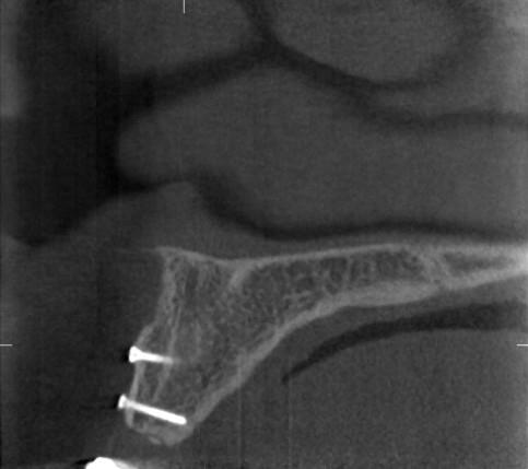
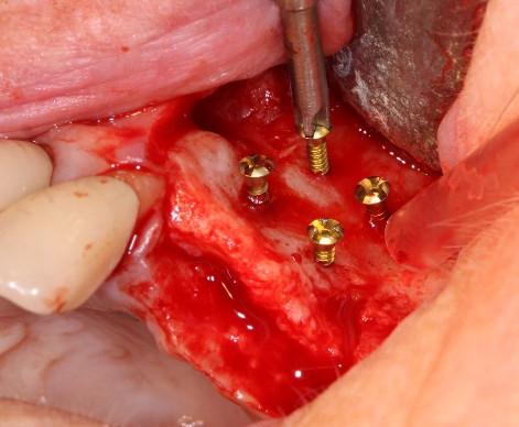
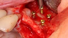
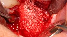
Lack of sagittal bone dimension will require buccal onlay augmentation, either with a mini-screw fixed autogenous bone block or with particulate bone substitute materials. Particulate bone may be kept in place using mini-screws as tent-poles, emerging 2-3 mm above the buccal bone plate. The latter procedure may occasionally be suitable even for the lower incisor region. Membranes are regularly used with success to cover osteotomies and graft materials. The implant is normally inserted according to a staged approach, i.e. 4-6 months post grafting.

Figure 1: Radiograph of buccal onlay block bone fixed with two mini-screws
Figures 2-3: Mini-screws used as tent-poles to keep bone particles in position


