-
0
Patient Assessment
- 0.1 Patient Demand
- 0.2 Anatomical location
-
0.3
Patient History
- 2.1 General patient history
- 2.2 Local history
-
0.4
Risk Assessment
- 3.1 Risk Assessment Overview
- 3.2 Age
- 3.3 Patient Compliance
- 3.4 Smoking
- 3.5 Drug Abuse
- 3.6 Recreational Drug and Alcohol Abuse
- 3.7 Condition of Natural Teeth
- 3.8 Parafunctions
- 3.9 Diabetes
- 3.10 Anticoagulants
- 3.11 Osteoporosis
- 3.12 Bisphosphonates
- 3.13 MRONJ
- 3.14 Steroids
- 3.15 Radiotherapy
- 3.16 Risk factors
-
1
Diagnostics
-
2
Treatment Options
-
2.1
Treatment planning
- 0.1 Non-implant based treatment options
- 0.2 Treatment planning conventional, model based, non-guided, semi-guided
- 0.3 Digital treatment planning
- 0.4 NobelClinician and digital workflow
- 0.5 Implant position considerations overview
- 0.6 Soft tissue condition and morphology
- 0.7 Site development, soft tissue management
- 0.8 Hard tissue and bone quality
- 0.9 Site development, hard tissue management
- 0.10 Time to function
- 0.11 Submerged vs non-submerged
- 0.12 Healed or fresh extraction socket
- 0.13 Screw-retained vs. cement-retained
- 0.14 Angulated Screw Channel system (ASC)
- 2.2 Treatment options esthetic zone
- 2.3 Treatment options posterior zone
- 2.4 Comprehensive treatment concepts
-
2.1
Treatment planning
-
3
Treatment Procedures
-
3.1
Treatment procedures general considerations
- 0.1 Anesthesia
- 0.2 peri-operative care
- 0.3 Flap- or flapless
- 0.4 Non-guided protocol
- 0.5 Semi-guided protocol
- 0.6 Guided protocol overview
- 0.7 Guided protocol NobelGuide
- 0.8 Parallel implant placement considerations
- 0.9 Tapered implant placement considerations
- 0.10 3D implant position
- 0.11 Implant insertion torque
- 0.12 Intra-operative complications
- 0.13 Impression procedures, digital impressions, intraoral scanning
- 3.2 Treatment procedures esthetic zone surgical
- 3.3 Treatment procedures esthetic zone prosthetic
- 3.4 Treatment procedures posterior zone surgical
- 3.5 Treatment procedures posterior zone prosthetic
-
3.1
Treatment procedures general considerations
-
4
Aftercare
Provisionalization procedures esthetic zone
Key points
- The provisional can be placed at various times.
- Provisionals must be well made and are the key to soft tissue health and for the development of the soft-tissue emergence profile.
- Provisional restorations serve as the template for the final restorations.
There are a variety of times that a provisional may be placed during the treatment phase. The classic approach is following the osseointegration phase and following the healing of the soft tissue subsequent to placing the healing abutment. The healing abutment is removed and the soft tissue evaluated for depth, tissue type, and architecture. The connection to the implant can either be with an abutment or directly to the implant via the restoration. The abutment / restoration must incorporate a non rotating feature to the implant connection. In the classic approach a either a provisional or final abutment may be placed followed by titanium provisional cylinder can be utilized and cut to length (assuming the access opening is in a favorable position) and the tooth contours developed to satisfy the esthetic and functional needs. This can also be accomplished by going directly to the implant platform with the provisional cylinder and following the same technique.
Another option would be to have indexed the implant position at the time of stage 1 surgery and have fabricated a master cast. The provisional can then be fabricated in the laboratory and placed with the permanent abutment (if desired) and a provisional. The advantage to this technique is that it allows the fabrication of a custom abutment with CAD CAM technology and gives many more options for materials. In patients with thin gingival biotype this may be very helpful since it allows the use of ceramic materials that are less likely to show through the soft tissue. One additional advantage of placing the abutment at this time is the potential for some soft tissue attachment exists. Thus by placing the final abutment and not having disturb the soft tissue more consistent results are possible.
The increasing acceptance of immediate placement and immediate load protocols has now allowed for the placement of the provisional at stage 1. Again the above mentioned options exist and with the ability to plan precise implant placement relative to adjacent structures (NobelClinician) also allows for the selection of the abutment. This enables the placement of the provisional simultaneously. The main concern then shifts to controlling the forces on the complex during the osseointegration phase. Patients need to be counseled on diet and understand fully the restrictions they must follow. This is also dependent on the implant reaching acceptable torque values thus insuring initial stability and proper healing. As cement may sometimes affect the healing if not completely removed it would be advisable to utilize screw retention when possible during this phase of treatment. One final advantage to this is the patient leaves with a tooth in place, not a healing abutment of cover screw. This could negate the need for a second surgery, and a patient having an improved quality of life with a tooth in place in the critical anterior zone.
In all instances there must be adequate time for the soft tissue to heal and mature prior to proceeding with impressions (whether traditional or digital) in order to achieve maximum esthetics. Once the patient and clinician are satisfied with provisional’s esthetics and soft tissue this information can be passed on to the dental laboratory via different recording mediums thus allowing the provisional to serve as a template of the final restoration.
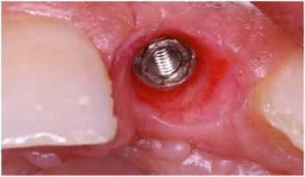
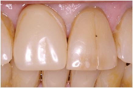
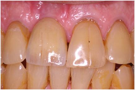
Figure 1 Figure 2
Single tooth immediate with provisional and ceramic abutment
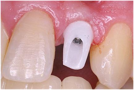
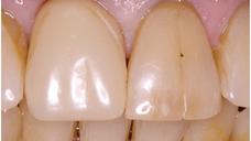
Figure 3: Ceramic abutment placed at stage 2 surgery 2wks post healing
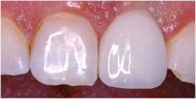
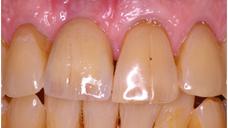
Figure 4 Figure 5
Single tooth provisional followed by final restoration notice how soft tissue was sculpted by the provisional
