-
0
Patient Assessment
- 0.1 Patient demand
- 0.2 Overarching considerations
- 0.3 Local history
- 0.4 Anatomical location
- 0.5 General patient history
-
0.6
Risk assessment & special high risk categories
- 5.1 Risk assessment & special high risk categories
- 5.2 age
- 5.3 Compliance
- 5.4 Smoking
- 5.5 Drug abuse
- 5.6 Recreational drugs and alcohol abuse
- 5.7 Parafunctions
- 5.8 Diabetes
- 5.9 Osteoporosis
- 5.10 Coagulation disorders and anticoagulant therapy
- 5.11 Steroids
- 5.12 Bisphosphonates
- 5.13 BRONJ / ARONJ
- 5.14 Radiotherapy
- 5.15 Risk factors
-
1
Diagnostics
-
1.1
Clinical Assessment
- 0.1 Lip line
- 0.2 Mouth opening
- 0.3 Vertical dimension
- 0.4 Maxillo-mandibular relationship
- 0.5 TMD
- 0.6 Existing prosthesis
- 0.7 Muco-gingival junction
- 0.8 Hyposalivation and Xerostomia
- 1.2 Clinical findings
-
1.3
Clinical diagnostic assessments
- 2.1 Microbiology
- 2.2 Salivary output
-
1.4
Diagnostic imaging
- 3.1 Imaging overview
- 3.2 Intraoral radiographs
- 3.3 Panoramic
- 3.4 CBCT
- 3.5 CT
- 1.5 Diagnostic prosthodontic guides
-
1.1
Clinical Assessment
-
2
Treatment Options
- 2.1 Mucosally-supported
-
2.2
Implant-retained/supported, general
- 1.1 Prosthodontic options overview
- 1.2 Number of implants maxilla and mandible
- 1.3 Time to function
- 1.4 Submerged or non-submerged
- 1.5 Soft tissue management
- 1.6 Hard tissue management, mandible
- 1.7 Hard tissue management, maxilla
- 1.8 Need for grafting
- 1.9 Healed vs fresh extraction socket
- 1.10 Digital treatment planning protocols
- 2.3 Implant prosthetics - removable
-
2.4
Implant prosthetics - fixed
- 2.5 Comprehensive treatment concepts
-
3
Treatment Procedures
-
3.1
Surgical
-
3.2
Removable prosthetics
-
3.3
Fixed prosthetics
-
3.1
Surgical
- 4 Aftercare
Panoramic radiographs
Key points
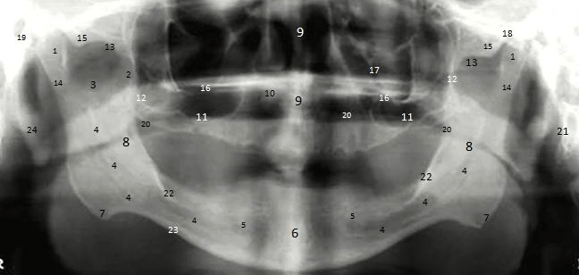
- Doctor should be familiar with recognizable anatomical structures
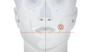
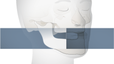
- The process of obtaining an excellent panoramic film is technique sensitive: head positioning, removal of facial/head jewelry etc.
- Digital radiography offers more options (documentation, measurements, exposure latitude)
Panoramic radiographs
When obtaining a panoramic film, one must be aware of the importance of following proper technique and carefully positioning the patient's skull between the X-ray generator and the film. Rotation of the film and radiation source often occurs along a sliding path and radiation exposure time varies between 5 and 20 seconds. Digital radiography offers more options and offers greater exposure latitude. However, panoramic films underestimate the lower jaw bone height and horizontal measurements are also not reliable. In addition, differences towards real measurements are more limited in the posterior areas.
Panoramic films have many positive elements and are widely used. They can reveal intra-osseous lesions or teeth and provide a screening tool for the maxillary sinus and orbit. Often overlooked due to a focus on maxillo-mandibular structures, arterial atheromas in the neck can be seen on panoramic films. In addition, and of practical clinical advantage, they are a useful alternative for patients with severe a gag reflex.
