-
0
Patient Assessment
- 0.1 Patient Demand
- 0.2 Anatomical location
-
0.3
Patient History
- 2.1 General patient history
- 2.2 Local history
-
0.4
Risk Assessment
- 3.1 Risk Assessment Overview
- 3.2 Age
- 3.3 Patient Compliance
- 3.4 Smoking
- 3.5 Drug Abuse
- 3.6 Recreational Drug and Alcohol Abuse
- 3.7 Condition of Natural Teeth
- 3.8 Parafunctions
- 3.9 Diabetes
- 3.10 Anticoagulants
- 3.11 Osteoporosis
- 3.12 Bisphosphonates
- 3.13 MRONJ
- 3.14 Steroids
- 3.15 Radiotherapy
- 3.16 Risk factors
-
1
Diagnostics
-
2
Treatment Options
-
2.1
Treatment planning
- 0.1 Non-implant based treatment options
- 0.2 Treatment planning conventional, model based, non-guided, semi-guided
- 0.3 Digital treatment planning
- 0.4 NobelClinician and digital workflow
- 0.5 Implant position considerations overview
- 0.6 Soft tissue condition and morphology
- 0.7 Site development, soft tissue management
- 0.8 Hard tissue and bone quality
- 0.9 Site development, hard tissue management
- 0.10 Time to function
- 0.11 Submerged vs non-submerged
- 0.12 Healed or fresh extraction socket
- 0.13 Screw-retained vs. cement-retained
- 0.14 Angulated Screw Channel system (ASC)
- 2.2 Treatment options esthetic zone
- 2.3 Treatment options posterior zone
- 2.4 Comprehensive treatment concepts
-
2.1
Treatment planning
-
3
Treatment Procedures
-
3.1
Treatment procedures general considerations
- 0.1 Anesthesia
- 0.2 peri-operative care
- 0.3 Flap- or flapless
- 0.4 Non-guided protocol
- 0.5 Semi-guided protocol
- 0.6 Guided protocol overview
- 0.7 Guided protocol NobelGuide
- 0.8 Parallel implant placement considerations
- 0.9 Tapered implant placement considerations
- 0.10 3D implant position
- 0.11 Implant insertion torque
- 0.12 Intra-operative complications
- 0.13 Impression procedures, digital impressions, intraoral scanning
- 3.2 Treatment procedures esthetic zone surgical
- 3.3 Treatment procedures esthetic zone prosthetic
- 3.4 Treatment procedures posterior zone surgical
- 3.5 Treatment procedures posterior zone prosthetic
-
3.1
Treatment procedures general considerations
-
4
Aftercare
インプラントのポジション(臼歯部領域)
Key points
- 近遠心空間(隣接歯間):7mm(→インプラント1本)、12mm(→インプラント2本)
- 口蓋頬側(Bucco-palatal)空間:6mm(通常インプラント: Ø 4mm)
- 隣接歯からの距離:≥1.5mm
- インプラント間空間:3mm.
- 上顎骨間(咬合)空間:>10mm.
- 解剖学的検討事項:上顎洞、下歯槽神経、オトガイ神経、リンガルコンキャビティ(下顎)
- 選択肢:可撤式部分義歯、樹脂接着の固定式補綴物、固定式部分義歯
解剖学的検討事項
臼歯部領域へのインプラント埋入に先立ち、パノラマX線写真および/または歯科CTもしくはCBCTスキャンによる詳細なX線写真検査の実施を推奨します。これにより、解剖学的な構造、骨質、神経血管等が視覚化されます。パノラマX線写真に関しては、拡大係数の補正を行う必要があります。インプラント先端と神経管との間の安全域2mmを順守する必要があります(Greensteinら。2008年)。
臨床面
臨床評価は、歯の間隙の近遠心空間(インプラント1本の場合は7mm以上、2本の場合は12mm以上)、口蓋頬側(bucco-palatal)空間(通常のインプラントを埋入できるよう6mm以上)、咬合面間の空間(固定式義歯の場合は6mm以上、可撤式義歯の場合は12mm以上)、増生を必要とする抜歯後の骨欠損、角化粘膜の幅、歯肉のバイオタイプ(薄いスキャロップ型か、厚く平坦か)、開口(>40mm)の測定から構成されます。
外科術式
インプラントの埋入は、タイミングを基準に即時、早期、遅延に分類されます。インプラントの即時埋入は、治療期間の短縮を始め、患者様にとっていくつか利点がありますが、常に実施できるとは限らず、またより高度な外科技術が求められます。初期固定は、臼歯の根間中隔(inter-radicular bony septum)にインプラントを埋入することにより、臼歯部領域でより容易に得ることができます。
細く鋭くとがった歯槽堤(インプラントの遅延埋入)は、事前の骨増生、または骨の高さが十分な場合は通常のサイズのインプラントに対し十分な幅を得るための歯槽頂部骨のプレパレーションがやむを得ず必要となることがあります。
インプラントのデザインには、頚部に加工を施したものと加工を施していないものがあります。これら2種類のデザインに関し、歯槽頂レベルに埋入した場合の骨吸収を比較した研究では、無負荷期間後に、頚部に加工を施していないインプラントに骨吸収がなかったことが示されました。しかし、歯槽頂部骨吸収は負荷後に生じることが多く、また歯周の観点からは、頚部に加工を施したインプラントの方がプラークを除去しやすいと考えられることから、頚部に加工がある否かの問題は、依然として議論が続いているという点について触れておく価値はあるでしょう。骨再形成は、歯槽頂上(epicrestal)に埋入される、頚部に加工を施したインプラント周囲で認められています。これらのインプラントを歯槽骨縁上(supracrestal)に埋入すると、骨吸収は減少する傾向にあります。
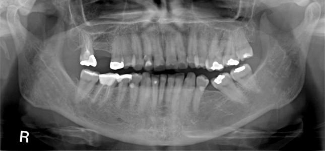
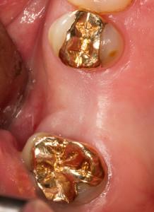
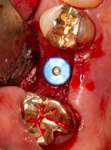
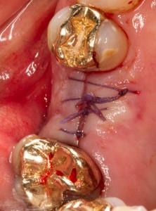
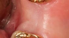
図1:喪失歯 #16 (#3 UNIV)
