-
0
Patient Assessment
- 0.1 Patient demand
- 0.2 Overarching considerations
- 0.3 Local history
- 0.4 Anatomical location
- 0.5 General patient history
-
0.6
Risk assessment & special high risk categories
- 5.1 Risk assessment & special high risk categories
- 5.2 age
- 5.3 Compliance
- 5.4 Smoking
- 5.5 Drug abuse
- 5.6 Recreational drugs and alcohol abuse
- 5.7 Parafunctions
- 5.8 Diabetes
- 5.9 Osteoporosis
- 5.10 Coagulation disorders and anticoagulant therapy
- 5.11 Steroids
- 5.12 Bisphosphonates
- 5.13 BRONJ / ARONJ
- 5.14 Radiotherapy
- 5.15 Risk factors
-
1
Diagnostics
-
1.1
Clinical Assessment
- 0.1 Lip line
- 0.2 Mouth opening
- 0.3 Vertical dimension
- 0.4 Maxillo-mandibular relationship
- 0.5 TMD
- 0.6 Existing prosthesis
- 0.7 Muco-gingival junction
- 0.8 Hyposalivation and Xerostomia
- 1.2 Clinical findings
-
1.3
Clinical diagnostic assessments
- 2.1 Microbiology
- 2.2 Salivary output
-
1.4
Diagnostic imaging
- 3.1 Imaging overview
- 3.2 Intraoral radiographs
- 3.3 Panoramic
- 3.4 CBCT
- 3.5 CT
- 1.5 Diagnostic prosthodontic guides
-
1.1
Clinical Assessment
-
2
Treatment Options
- 2.1 Mucosally-supported
-
2.2
Implant-retained/supported, general
- 1.1 Prosthodontic options overview
- 1.2 Number of implants maxilla and mandible
- 1.3 Time to function
- 1.4 Submerged or non-submerged
- 1.5 Soft tissue management
- 1.6 Hard tissue management, mandible
- 1.7 Hard tissue management, maxilla
- 1.8 Need for grafting
- 1.9 Healed vs fresh extraction socket
- 1.10 Digital treatment planning protocols
- 2.3 Implant prosthetics - removable
-
2.4
Implant prosthetics - fixed
- 2.5 Comprehensive treatment concepts
-
3
Treatment Procedures
-
3.1
Surgical
-
3.2
Removable prosthetics
-
3.3
Fixed prosthetics
-
3.1
Surgical
- 4 Aftercare
Splints for imaging and surgery
Key points
- Based on the diagnostic provisional set-up, planning tools (radiographic templates), surgery tools (surgical templates) and temporary dentures can be fabricated
- Radiographic/imaging templates help calculate available vertical height of the jaws and possible depth of surgical drill penetration, and help indicate favorable implant positions
- Prosthodontically driven implant treatment uses a clear resin duplicate of the laboratory teeth set-up, as a surgical template
Diagnostic and planning guides
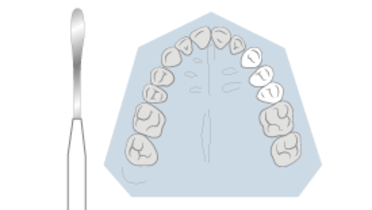
The primary goal of creating a preliminary teeth arrangement for a try-in diagnostic evaluation is the opportunity to visualize those specific morphological features that impact functional and esthetic considerations for each treated patient. This trial diagnostic set-up is also used to plan, as well as guide the overlapping selection of both implant locations and their subsequent surgical placement.
The teeth arrangement in the set-up becomes the basis for a radiographic template, fabrication of a surgical guide, and if necessary the making of suitably designed interim complete dentures.
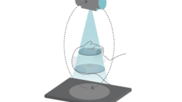
Diagnostic imaging is inarguably essential for the selection of host bone sites for implant placement. The imaging prescription is based on the clinical examination, a determination of the type of information required, and an evaluation of the radiation risk - benefit ratio for the patient.
Imaging templates
The objective of an radiographic guide is to visualize the bone anatomy in relation to the ideal final restoration. Simple radiographic/imaging templates with metallic markers of known diameters, placed in canine positions, help calculate available vertical height of the jaw bones. The template helps indicate favorable implant positions and may also provide approximate preliminary calculation of possible depth of surgical drill penetration. The comparison of the actual size of the markers with their size on the radiographs allows to calculate the distortion factor of the X-ray and to determine the distances of important anatomic structures. Particularly accurate measurement of bone dimensions and their transfer to the surgical site can also be achieved if radiopaque markers are incorporated in the regions of potential implant sites in a tomographic image, including the angulation at which the implant will be placed in the bone. This eliminates or minimizes any guesswork related to the orientation of the bone measurements.
Surgical templates / guides
Prosthetically driven implant treatment uses a clear resin duplicate of the laboratory teeth set-up. Radiopaque titanium pins can be additionally integrated into this template to clearly mark selected teeth positions. This is also possible via a laboratory made surgical splint.
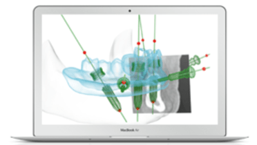
A computer-assisted planning program may also be selected to transfer the analog tool into digital data obtained by means of digital radiographic technology. Computer-assisted implant planning allows for a virtual evaluation of the relation between jawbone and denture parameters in all 3 dimensions. The fabrication of a surgical splint for a fully guided surgical protocol is also based on these software data.


