-
0
Patient Assessment
- 0.1 Patient Demand
- 0.2 Anatomical location
-
0.3
Patient History
- 2.1 General patient history
- 2.2 Local history
-
0.4
Risk Assessment
- 3.1 Risk Assessment Overview
- 3.2 Age
- 3.3 Patient Compliance
- 3.4 Smoking
- 3.5 Drug Abuse
- 3.6 Recreational Drug and Alcohol Abuse
- 3.7 Condition of Natural Teeth
- 3.8 Parafunctions
- 3.9 Diabetes
- 3.10 Anticoagulants
- 3.11 Osteoporosis
- 3.12 Bisphosphonates
- 3.13 MRONJ
- 3.14 Steroids
- 3.15 Radiotherapy
- 3.16 Risk factors
-
1
Diagnostics
-
2
Treatment Options
-
2.1
Treatment planning
- 0.1 Non-implant based treatment options
- 0.2 Treatment planning conventional, model based, non-guided, semi-guided
- 0.3 Digital treatment planning
- 0.4 NobelClinician and digital workflow
- 0.5 Implant position considerations overview
- 0.6 Soft tissue condition and morphology
- 0.7 Site development, soft tissue management
- 0.8 Hard tissue and bone quality
- 0.9 Site development, hard tissue management
- 0.10 Time to function
- 0.11 Submerged vs non-submerged
- 0.12 Healed or fresh extraction socket
- 0.13 Screw-retained vs. cement-retained
- 0.14 Angulated Screw Channel system (ASC)
- 2.2 Treatment options esthetic zone
- 2.3 Treatment options posterior zone
- 2.4 Comprehensive treatment concepts
-
2.1
Treatment planning
-
3
Treatment Procedures
-
3.1
Treatment procedures general considerations
- 0.1 Anesthesia
- 0.2 peri-operative care
- 0.3 Flap- or flapless
- 0.4 Non-guided protocol
- 0.5 Semi-guided protocol
- 0.6 Guided protocol overview
- 0.7 Guided protocol NobelGuide
- 0.8 Parallel implant placement considerations
- 0.9 Tapered implant placement considerations
- 0.10 3D implant position
- 0.11 Implant insertion torque
- 0.12 Intra-operative complications
- 0.13 Impression procedures, digital impressions, intraoral scanning
- 3.2 Treatment procedures esthetic zone surgical
- 3.3 Treatment procedures esthetic zone prosthetic
- 3.4 Treatment procedures posterior zone surgical
- 3.5 Treatment procedures posterior zone prosthetic
-
3.1
Treatment procedures general considerations
-
4
Aftercare
Diagnostic casts and wax-up
Key points
- Accurate diagnostic casts and an inter-occlusal/arch registration permits precise evaluation of existing morphological determinants and treatment possibilities.
- A diagnostic wax-up is a dental diagnostic procedure in which planned restorations are developed in wax on a diagnostic cast to determine optimal clinical and laboratory procedures necessary to achieve the desired esthetics and function.
Diagnostic casts and wax-up
It is important to diagnose available space and occlusion for either the implant-supported single-tooth restoration or a fixed denture. Diagnostic casts and a diagnostic wax-up will enhance the predictability of the treatment.
The diagnostic wax-up is a tool that can be used for diagnosis and treatment of single-tooth implants , using wax or denture teeth set in wax. This tool provides important diagnostic information that will indicate whether a specific treatment is appropriate. It also assists in the selection of a proper restoration method and may indicate the need for orthodontic treatment or preprosthetic surgery. The diagnostic casts and wax-up can assist in estimating the amount of restorative space available and point out any need for treatment in the opposing arch to obtain such space. It can help in evaluating the planned occlusal scheme and indicate which modifications are needed in the remaining dentition. Additionally, the diagnostic wax-up can be used as means of communication between the clinician, the technician, and the patient.
- The following steps are part of the diagnostic casts and wax-up procedure:
- alginate impressions, occlusal records, if necessary a set of photographs
- fabrication of two sets of study models, mounted on an articulator (one to keep the original situation, one to use for the wax-up);
- diagnostic wax-up;
- communication between the clinician, the technician, and the patient illustrating the plan three-dimensionally;
- if necessary, adjustments to the initial plan;
- if necessary, dependent on the extensiveness of the treatment plan, a mock-up of autopolymerizing resin can be made indirectly by the laboratory to be evaluated intraorally.
The diagnostic wax-up can also be used as a treatment tool. In edentulous spaces, it can be used to form radiographic and surgical implant placement guides.
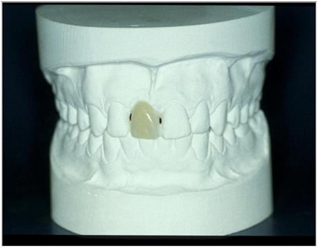
Figure 1: Diagnostic casts, missing tooth in position 11 (#8 UNIV) with diagnostic wax-up; no further restorative treatment necessary.
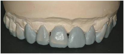
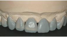
Figure 2: Diagnostic cast, missing tooth in position 21 (#9 UNIV) with diagnostic wax-up; extensive further restorative treatment necessary.


