-
0
Patient Assessment
- 0.1 Patient Demand
- 0.2 Anatomical location
-
0.3
Patient History
- 2.1 General patient history
- 2.2 Local history
-
0.4
Risk Assessment
- 3.1 Risk Assessment Overview
- 3.2 Age
- 3.3 Patient Compliance
- 3.4 Smoking
- 3.5 Drug Abuse
- 3.6 Recreational Drug and Alcohol Abuse
- 3.7 Condition of Natural Teeth
- 3.8 Parafunctions
- 3.9 Diabetes
- 3.10 Anticoagulants
- 3.11 Osteoporosis
- 3.12 Bisphosphonates
- 3.13 MRONJ
- 3.14 Steroids
- 3.15 Radiotherapy
- 3.16 Risk factors
-
1
Diagnostics
-
2
Treatment Options
-
2.1
Treatment planning
- 0.1 Non-implant based treatment options
- 0.2 Treatment planning conventional, model based, non-guided, semi-guided
- 0.3 Digital treatment planning
- 0.4 NobelClinician and digital workflow
- 0.5 Implant position considerations overview
- 0.6 Soft tissue condition and morphology
- 0.7 Site development, soft tissue management
- 0.8 Hard tissue and bone quality
- 0.9 Site development, hard tissue management
- 0.10 Time to function
- 0.11 Submerged vs non-submerged
- 0.12 Healed or fresh extraction socket
- 0.13 Screw-retained vs. cement-retained
- 0.14 Angulated Screw Channel system (ASC)
- 2.2 Treatment options esthetic zone
- 2.3 Treatment options posterior zone
- 2.4 Comprehensive treatment concepts
-
2.1
Treatment planning
-
3
Treatment Procedures
-
3.1
Treatment procedures general considerations
- 0.1 Anesthesia
- 0.2 peri-operative care
- 0.3 Flap- or flapless
- 0.4 Non-guided protocol
- 0.5 Semi-guided protocol
- 0.6 Guided protocol overview
- 0.7 Guided protocol NobelGuide
- 0.8 Parallel implant placement considerations
- 0.9 Tapered implant placement considerations
- 0.10 3D implant position
- 0.11 Implant insertion torque
- 0.12 Intra-operative complications
- 0.13 Impression procedures, digital impressions, intraoral scanning
- 3.2 Treatment procedures esthetic zone surgical
- 3.3 Treatment procedures esthetic zone prosthetic
- 3.4 Treatment procedures posterior zone surgical
- 3.5 Treatment procedures posterior zone prosthetic
-
3.1
Treatment procedures general considerations
-
4
Aftercare
Panoramic
Key points
- Panoramic radiograph depicts a two-dimensional view of complicated three-dimensional structures.
- Vertical height of bone can be assessed with panoramic radiograph.
- It can be supplemented with intraoral radiographs.
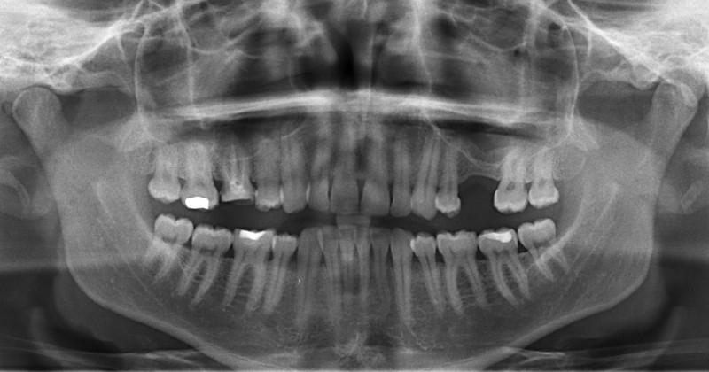
Panoramic radiograph still remain the most popular and widely available diagnostic modality in dentistry. Basic anatomy of the jaws and any related pathologic findings can be evaluated with convenience. Panoramic radiograph is easy to perform and produces a single image of the mandible, maxilla, teeth, temporomandibular joints and the lower half of the maxillary sinuses. Nevertheless, panoramic radiograph depicts only a two-dimensional view of complicated three-dimensional sections of the jaws of variable thickness.
This technique features many potential intrinsic mistakes, not least in patient positioning, ranging from variable magnification factor, overlapping of the image at the premolar region, superimposition of the cervical spine in the incisor region, and presence of hard tissue and soft tissue artifacts.

Figure 1: panoramic radiograph.
During the exposure the x-ray source and detector rotate synchronously around the patient producing a curved plane tomography. The focal trough is fixed relative to the X-ray tube and the detector. The patient's jaws should be positioned accurately within this layer that is considerably thinner in the anterior region than the posterior regions. Therefore, both regions can become distorted and thus not represent the jaw dimensions correctly. Despite this limitation, panoramic radiographs can still be used in implant dentistry to provide a quick estimate of the bone height by taking the magnification into account. Magnification may vary between different types of panoramic radiographs and particularly affects measurements made in a horizontal direction.
The main advantage of the panoramic radiograph remain its ability to detect possible pathology within the hard tissue, as well as, to assess patient eligibility for treatment. Also, the panoramic radiograph, together with intraoral images, may be the preferred exam to be done during follow-up examinations. However, dentomaxillofacial imaging, especially for diagnostic and planning purposes, continues the move towards 3D imaging.
Visit the treatment guidelines section for edentulous (treatment-guidelines/edentulous/diagnostics/diagnostic-imaging/panoramic) to see the complete anatomical landmarks visualized on a panoramic radiograph.
