-
0
Patient Assessment
- 0.1 Patient Demand
- 0.2 Anatomical location
-
0.3
Patient History
- 2.1 General patient history
- 2.2 Local history
-
0.4
Risk Assessment
- 3.1 Risk Assessment Overview
- 3.2 Age
- 3.3 Patient Compliance
- 3.4 Smoking
- 3.5 Drug Abuse
- 3.6 Recreational Drug and Alcohol Abuse
- 3.7 Condition of Natural Teeth
- 3.8 Parafunctions
- 3.9 Diabetes
- 3.10 Anticoagulants
- 3.11 Osteoporosis
- 3.12 Bisphosphonates
- 3.13 MRONJ
- 3.14 Steroids
- 3.15 Radiotherapy
- 3.16 Risk factors
-
1
Diagnostics
-
2
Treatment Options
-
2.1
Treatment planning
- 0.1 Non-implant based treatment options
- 0.2 Treatment planning conventional, model based, non-guided, semi-guided
- 0.3 Digital treatment planning
- 0.4 NobelClinician and digital workflow
- 0.5 Implant position considerations overview
- 0.6 Soft tissue condition and morphology
- 0.7 Site development, soft tissue management
- 0.8 Hard tissue and bone quality
- 0.9 Site development, hard tissue management
- 0.10 Time to function
- 0.11 Submerged vs non-submerged
- 0.12 Healed or fresh extraction socket
- 0.13 Screw-retained vs. cement-retained
- 0.14 Angulated Screw Channel system (ASC)
- 2.2 Treatment options esthetic zone
- 2.3 Treatment options posterior zone
- 2.4 Comprehensive treatment concepts
-
2.1
Treatment planning
-
3
Treatment Procedures
-
3.1
Treatment procedures general considerations
- 0.1 Anesthesia
- 0.2 peri-operative care
- 0.3 Flap- or flapless
- 0.4 Non-guided protocol
- 0.5 Semi-guided protocol
- 0.6 Guided protocol overview
- 0.7 Guided protocol NobelGuide
- 0.8 Parallel implant placement considerations
- 0.9 Tapered implant placement considerations
- 0.10 3D implant position
- 0.11 Implant insertion torque
- 0.12 Intra-operative complications
- 0.13 Impression procedures, digital impressions, intraoral scanning
- 3.2 Treatment procedures esthetic zone surgical
- 3.3 Treatment procedures esthetic zone prosthetic
- 3.4 Treatment procedures posterior zone surgical
- 3.5 Treatment procedures posterior zone prosthetic
-
3.1
Treatment procedures general considerations
-
4
Aftercare
Occlusion and functional aspects
Key points
- Existing occlusion is an important part of pre-treatment diagnostics.
- In (partially) dentate patients most functional tooth contacts occur in a mandibular position slightly anterior to Centric Relation (CR), a position known as Centric Occlusion (CO).
- Occlusion must give the possibility to design a future implant-supported restoration without high non-axial forces on the occlusal plane.
Occlusion
Occlusion describes the relationship between the opposing masticating surfaces of teeth and the movements of the mandible dictated by way of the temporomandibular joint and associated orofacial musculature.
In dentate patients most functional tooth contacts occur in a mandibular position slightly anterior to Centric Relation (CR), a position known as Centric Occlusion (CO). This position can be recorded via one of the several registration methods and materials. Recording of patient-specific, vertical and horizontal jaw relations is a necessary starting point when planning for occlusal rehabilitation. The optimum occlusal scheme is mutual protection, in which the posterior teeth contact simultaneously and equally in CO, the canines disclude the posterior teeth in lateral excursions, and the anterior teeth disclude the posterior teeth in protrusion.
Although rehabilitation with an implant-supported single-tooth restoration concerns only a small part of the total occlusal plane, it is important to diagnose the type of occlusion and deviations. All restorative dentistry is affected by occlusal forces on the teeth in function. A single-tooth implant-supported restoration must be protected to prevent biological and technical complications, occlusion must give the possibility to design a future restoration without high non-axial forces on the occlusal plane.
The mounting of models to aid in diagnosis and treatment planning is invaluable especially where multiple restorations are planned. Virtual planning on models by way of adjustments or wax-ups provides the clinician with foresight as to the achievability and predictability of a chosen plan.
If necessary, one should consider to reorganize the occlusion before dental implant treatment; not only for protection of the implant and its crown, but because of the benefit for the whole dentition. Sometimes adjusting of the palatal surfaces of the 13 and 23 is enough to steepen the canine guidance and improve separation of the posterior teeth in lateral excursions. In this way, non-axial forces can be minimized.

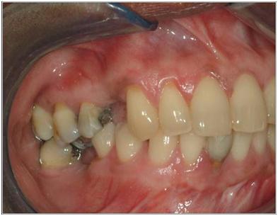
Figure1

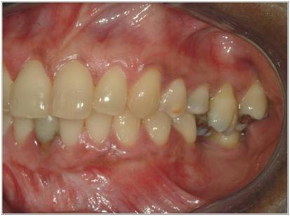
Figure 2
Patient with a wish for implant-supported restorations; multiple occlusal deviations, need for diagnostic models and wax-up and possibly the need for reorganizing of occlusion in advance of dental implant treatment.
