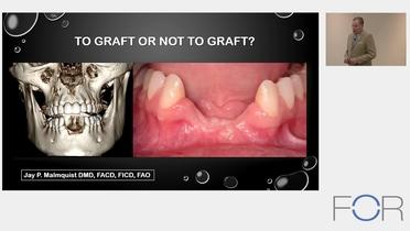
Grafting or Graftless - different treatment approaches for single teeth
Video highlights
- Graftless treatment options for single tooth replacements
- Sequence of grafting and implant placement
- Single tooth replacement in anterior and posterior regions
In the first part of our 3-part series “To graft or not to graft”, FOR Experts Dr. Edith Groenendijk, Dr. Ana Ferro and Dr. Jay Malmquist discuss the treatment options for missing teeth with and without grafting procedures.
Dr. Edith Groenendijk, who runs a practice in The Hague, Netherlands, opens the session focusing the discussion on single tooth treatments in the aesthetic area. Preferring a minimally invasive approach, She observes that single tooth replacements can be very challenging, especially when the three-dimensional biology is not fully understood. Even the slightest mistake during diagnosis of the indication can lead to undesirable aesthetic outcomes. Furthermore, suboptimal decisions on the surgical and prosthetic approach can dramatically impact the aesthetic results. The resulting poor aesthetic outcomes are often difficult to repair and restore. Dr. Groenendijk asks the audience to think about what happens when a tooth is extracted without treatment of the alveolar socket. Scientific data shows a dramatic alveolar bone loss of up to 63% can be expected within the first month. This is a significant amount and these missing tissues need to be repaired.
In a case presentation, the site is opened following an early implant placement protocol and a V-shaped defect is observed. An implant is placed into the one-wall defect with bone grafting material in front of the implant. Dr. Groenendijk asks the audience, “Is this a predictable?”. The audience agrees with her that this a predictable as the results can be predicted. But, it will not be an optimal result as the implant is placed exactly in the place where the bone growth is expected – bone grows from bone and not from implant. She shares her experience with the application of early implant placement, as introduced by Daniel Buser in 2004 and how buccal concavity was a common outcome. Dr. Groenendijk credits Dr. Ueli Grunder in her subsequent treatment approach to build a hard tissue graft of minimum 2mm in front of the implant to stabilize the soft tissue for the long term. Dr. Groenendijk recommends an implant therapy in the aesthetic zone should either be immediate or delayed.
Dr. Groenendijk presents a 45 years old female, who presented herself to the office with the request to get a new crown on a central incisor. After reviewing the CBCT, Dr. Groenendijk informed the patient that the tooth had a horizontal fracture, a massive bone loss and an inflammation was present. There was already some soft tissue recession present and the distal papilla was also compromised. After presenting the initial situation, she asks the audience to select from the following three treatment options:
- Option: follow a delayed protocol and graft
- Option: follow a delayed protocol and preserve
- Option: follow an immediate protocol and preserve
The audience is indecisive between the first and third option. This aligns with the literature as there is no consensus on how to treat this situation. Dr. Groenendijk shares her treatment approach: immediate implant placement. Her rationale: as long as bony ingrowth from the interdental septa and the cranial can be expected based on the pre- operative CBCT scans, she places an immediate implant followed by a ridge preservation procedure in front of the implant. And, as long as the interdental septa are intact, papilla ingrowth is very predictable because when the distance between the bone crest and the contact point does not exceed 5 millimeters. She explains the implant position, being maximal to the palate, and the socket itself is used for a bone graft, i.e. bone preservation. Showing a 1-year follow up of the case, she mentioned the patient’s focus of was not on the distal papilla, which was already compromised at initial presentation and could not be restored 100%. It is important to discuss this with the patient before the treatment is started. Dr. Groenendijk summarized her procedure so that after tooth extraction, a preparation is made maximal to the palatal, if possible, into the palatal wall. About 5mms of bone height is needed in the palato-apical area to reach primary stability, and the implant is restored directly thereafter. Firstly, the socket is filled with bone substitute, which is considered to be the ridge preservation procedure and then the implant is installed. The socket is simply closed by the creation of a composite crown on top. No membrane is used, no connective tissue graft, and no flap is raised. The experienced results are supported by the scientific literature showing immediate implant placement often is associated with soft tissue recessions. When explaining why, she looked at the three-dimensional biology, were thick buccal crests are rare, with only 3% of patients presenting with a buccal crest of more than 2mms. A thick buccal crest is not expected to resorb significantly post-extraction. The majority of patients show a thin buccal crest with less than 1mm of thickness. This bony aspect consists of bundle bone and is vascularized by the periodontal ligamentum. With the extraction of a tooth, this blood supply is taken away which leads to its destruction. The literature confirms a vertical crestal bone loss of 7.5mm within eight weeks post-extraction can be expected. And when such a case is opened up, a V-shaped defect is present. So intact sockets are bone defects after eight weeks. Fresh extracted sockets also form an ideal situation for preservation of the alveolar volume because bone ingrowth from the buccal side is not expected. A nice four wall defect is present which is predictable to graft and directly after extraction growth factors and regenerative cells are present, which is very favorable for wound healing. By placing the implants maximal to the palatal, sufficient space is present for new bone ingrowth in front of the implants. To validate this method, a prospective clinical case series including 100 patients with both intact socket as buccal bone defects was started. The investigators found a tremendous improvement of the soft tissues from a PES score of 9.9 towards 12 after one year post surgery. Stable after three years, ist was also found was that bone defect didn't influence the soft tissue aesthetic outcome. This could be achieved because in those situations it is possible to overcontour the alveolar process. This leads to the assumption that is an advantage when the buccal crest is not present because that dimension is lost anyways. Dr. Groenendijk continues by displaying several cases showing the development of the soft tissue over several years. She concludes with a statement about her treatment approach: prevention is better than healing and that preservation is preferred over grafting, before introducing the next speaker.
Dr. Ana Ferro opens her presentation by thanking Dr. Groenendijk for her research in the matter and that it is important to develop protocols. Protocols help to develop a routine process which helps to compare cases and draw conclusions. Although the treatment of full arch cases is Dr. Ferro’s specialty, she also treats other indications. When planning and treating a case, she reminds us to keep the patient in mind and include them in the conversation to ensure their needs are addressed. She illustrates her treatment approach with a pie graph showing equal sections for the patient, the surgeon and materials. The patient comes with a diagnosis, desires and needs. The surgeon brings his/her knowledge, skills and experience and the materials represent the tools and products available. Presenting her first case, a 32-year-old female was in need of a treatment of the first upper molar. The patient, who was also a friend, had high aesthetic demands. During the anamnesis, Dr. Ferro noticed that the tooth already had furcation involvement in terms of sinus symptoms and gum recession. The patient complained about food impaction and a slight mobility was noticeable. The CBCT showed the need to extract the tooth. After the extraction, there would not be enough bone to place an implant, so Dr. Ferro was aware that a sinus lift procedure was necessary. She intensely discussed and defined several treatment options with the patient. Presenting these options, Dr. Ferro asks the audience to select their preferred treatment:
- Option: extraction of the tooth, after 4 months perform a sinus lift, after 6 months place implant, after 6 months place final crown
- Option: extraction of the tooth, after 4 months perform a sinus lift, after 6 months place implant with immediate crown
- Option: extraction of tooth and perform sinus lift, after 6 months place implant with immediate crown
- Option: perform sinus lift, after 6 months extraction of tooth, implant placement and immediate crown
The audience showed a clear preference for options 1 and 4. Dr. Ferro considers the patient demand to never be without tooth during the treatment – even for the molar. She also highlights that the treatment decision should be a shared responsibility between the surgeon and the patient. After considering other critical aspects, such as any medical history regarding the sinus, does the tooth need periodontal treatment, is it stable despite the furcation? Moving on to the treatment, Dr. Ferro explained that she performed the sinus lift using xenograft material with a lateral window approach. When moving on to extracting the tooth, she ensured that it was gently removed, ensuring that the bone between the tooth roots was kept intact. She concludes the case presentation with a photo her friend sent her some weeks ago, which shows a 11-year follow-up for this treatment.
References
[1] Tan WL, Wong TL, Wong MC, Lang NP. A systematic review of post-extractional alveolar hard and soft tissue dimensional changes in humans. Clin Oral Implants Res. 2012 Feb;23 Suppl 5:1-21. doi: 10.1111/j.1600-0501.2011.02375.x. PMID: 22211303.
[2] Groenendijk E, Bronkhorst EM, Meijer GJ. Does the pre-operative buccal soft tissue level at teeth or gingival phenotype dictate the aesthetic outcome after flapless immediate implant placement and provisionalization? Analysis of a prospective clinical case series. Int J Implant Dent. 2021 Aug 27;7(1):84. doi: 10.1186/s40729-021-00366-3. PMID: 34448101; PMCID: PMC8390706.
[3] Groenendijk E, Staas TA, Bronkhorst EM, Raghoebar GM, Meijer GJ. Factors Associated with Esthetic Outcomes of Flapless Immediate Placed and Loaded Implants in the Maxillary Incisor Region-Three-Year Results of a Prospective Case Series. J Clin Med. 2023 Mar 31;12(7):2625. doi: 10.3390/jcm12072625. PMID: 37048707; PMCID: PMC10094793.
[4] Huynh-Ba G, Pjetursson BE, Sanz M, Cecchinato D, Ferrus J, Lindhe J, Lang NP. Analysis of the socket bone wall dimensions in the upper maxilla in relation to immediate implant placement. Clin Oral Implants Res. 2010 Jan;21(1):37-42. doi: 10.1111/j.1600-0501.2009.01870.x. PMID: 20070745.
[5] Araújo MG, Lindhe J. Dimensional ridge alterations following tooth extraction. An experimental study in the dog. J Clin Periodontol. 2005 Feb;32(2):212-8. doi: 10.1111/j.1600-051X.2005.00642.x. PMID: 15691354.
[6] Chappuis V, Engel O, Reyes M, Shahim K, Nolte LP, Buser D. Ridge alterations post-extraction in the esthetic zone: a 3D analysis with CBCT. J Dent Res. 2013 Dec;92(12 Suppl):195S-201S. doi: 10.1177/0022034513506713. Epub 2013 Oct 24. PMID: 24158340; PMCID: PMC3860068.
[7] Grunder U, Gracis S, Capelli M. Influence of the 3-D bone-to-implant relationship on esthetics. Int J Periodontics Restorative Dent. 2005 Apr;25(2):113-9. PMID: 15839587.
[8] Qahash M, Susin C, Polimeni G, Hall J, Wikesjö UM. Bone healing dynamics at buccal peri-implant sites. Clin Oral Implants Res. 2008 Feb;19(2):166-72. doi: 10.1111/j.1600-0501.2007.01428.x. Epub 2007 Nov 26. PMID: 18039337.
[9] Groenendijk E, Staas TA, Bronkhorst E, Raghoebar GM, Meijer GJ. Immediate implant placement and provisionalization: Aesthetic outcome 1 year after implant placement. A prospective clinical multicenter study. Clin Implant Dent Relat Res. 2020 Apr;22(2):193-200. doi: 10.1111/cid.12883. Epub 2020 Jan 28. PMID: 31991527.
[10] Groenendijk E, Staas TA, Bronkhorst EM, Raghoebar GM, Meijer GJ. Factors Associated with Esthetic Outcomes of Flapless Immediate Placed and Loaded Implants in the Maxillary Incisor Region-Three-Year Results of a Prospective Case Series. J Clin Med. 2023 Mar 31;12(7):2625. doi: 10.3390/jcm12072625. PMID: 37048707; PMCID: PMC10094793.
[11] Testori T, Weinstein T, Taschieri S, Wallace SS. Risk factors in lateral window sinus elevation surgery. Periodontol 2000. 2019 Oct;81(1):91-123. doi: 10.1111/prd.12286. PMID: 31407430.
Clinical topics
Additional resources
Questions
Ask a questionIs it really professional?
In this presentation, Dr Edith Groenendijk presented a case that I published on the FOR website in 2014. First I am annoyed because she did without having the curtesy to ask for my permission even though she wrote on the slide that I gave it, and second because she did with a complete misunderstanding of the case, putting my name upfront with false ideas of how I treat my patients. This is clearly not professional to denigrate treatment options of others and even more without showing the full scope of the case. I feel she could take her own cases to criticize in a public presentation. I would be happy to discuss why I treated this patient this way and the 10-year outcome, but definitely not this way.
This is definitely not the spirit in which I signed up and agreed to publish my cases on FOR.
Well-respected colleague,
I am truly sorry that you feel offended by my presentation, this wasn't my goal.
Actually, I did ask for your permission to use this case, and I already send the proof of your approval to you personally. Nevertheless, now you have objections, I will not use the case anymore.
In my presentation, I do not state that you did bad work (I even say that I could not have done it better, ending up with buccal concavities myself all the time using this procedure). I only illustrated that there is a high risk of postoperative buccal concavities using an early placement procedure.
I invite you to discuss this in peace and privately, and hope I can buy you a beer at a future congress...
Best regards, Edith


When u have a situation where Posterior Maxillary region only 6mm of Bone Height ( Bilateral). Is it ok to go for ISL or DSL?
In reply to When u have a situation where Posterior Maxillary region only 6mm of Bone Height ( Bilateral). Is it ok to go for ISL or DSL? by Anonymous
ISL
In reply to When u have a situation where Posterior Maxillary region only 6mm of Bone Height ( Bilateral). Is it ok to go for ISL or DSL? by Anonymous
Thank you