The Nightmare of Peri-Implant Papilla
Clinical Challenge
Replacing two adjacent teeth in the anterior maxilla represents one of the most complex challenges in aesthetic implant dentistry, particularly when peri-implant papilla preservation is the primary objective. This clinical case involves a 36-year-old female patient who suffered dental trauma resulting in infraosseous root fractures of the maxillary canine and lateral incisor, combined with very high aesthetic expectations due to a high smile line and a gummy smile.
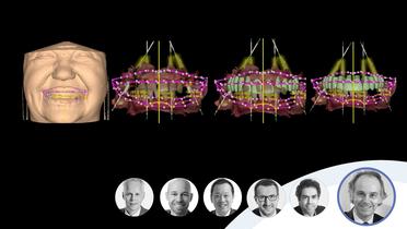
The treatment strategy was designed to achieve optimal soft tissue aesthetics while minimizing surgical invasiveness. Bone reconstruction procedures were intentionally avoided, and the patient’s request for a fixed provisional restoration throughout the entire treatment was respected. A fully digital workflow was adopted, integrating orthodontic, surgical, and prosthetic phases to preserve peri-implant tissues and restore the pre-trauma gingival architecture, with particular focus on interimplant papilla reconstruction and canine eminence restitution.
Evaluation & Diagnosis
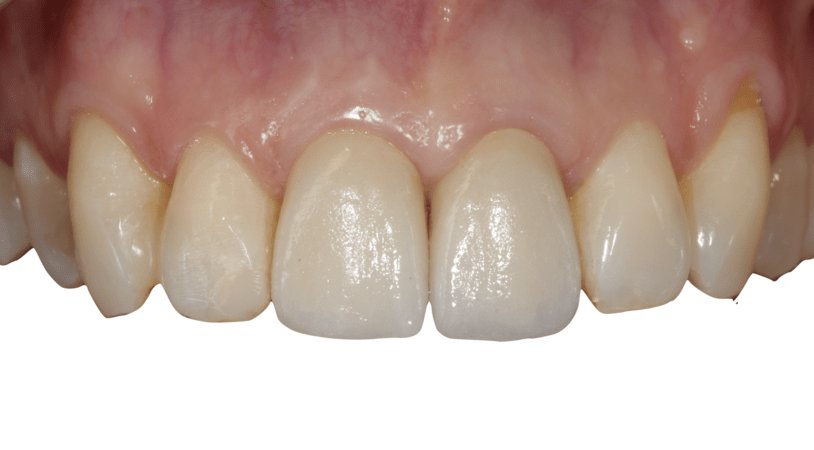
Baseline intraoral frontal view
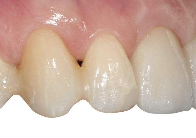
Preoperative clinical view of upper maxilla
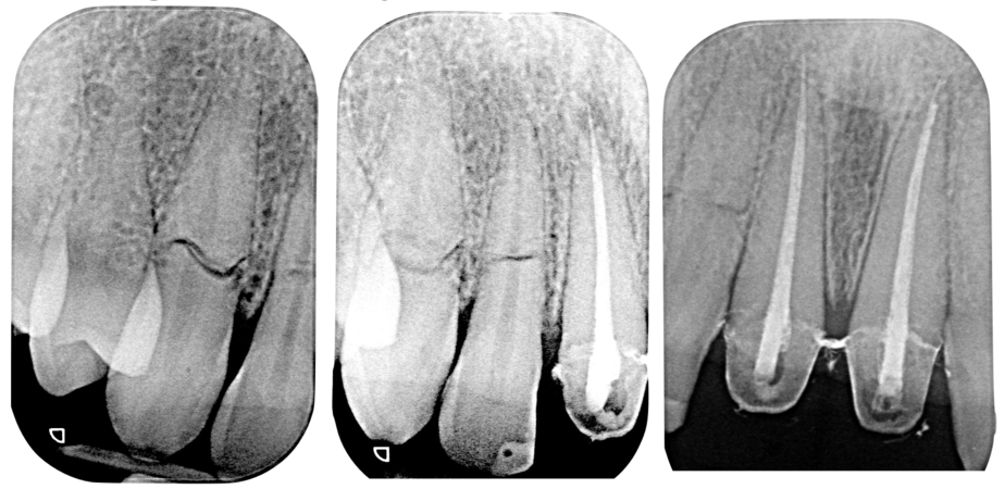
Horizontal root fractures at 1.3 and 1.2; periapical radiolucency at 1.1
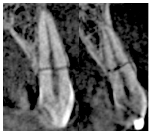
Root fractures
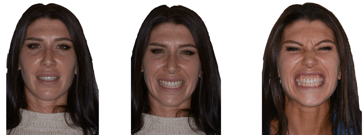
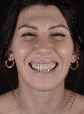
Baseline, natural smile
Clinical and radiographic examination revealed non-restorable infraosseous root fractures of teeth 1.3 and 1.2, associated with pulp necrosis of the maxillary central incisors. The patient presented a thin periodontal biotype, high smile line, and gummy smile, all of which significantly increased the aesthetic risk of implant rehabilitation in the anterior zone. The need to replace two adjacent teeth further amplified the challenge of maintaining soft tissue volume and papilla height.
The case was classified as a multi-unit treatment in the aesthetic zone of the anterior maxilla. Digital planning allowed precise assessment of hard and soft tissue conditions and facilitated a minimally invasive strategy focused on tissue preservation. The diagnostic phase confirmed that a combined orthodontic and surgical approach would be necessary to optimize tissue conditions prior to implant placement and soft tissue augmentation.
Progress & Completion
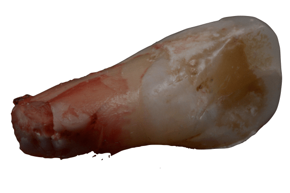
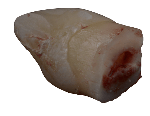
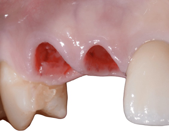
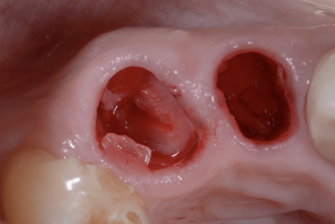
Extraction of coronal fragments
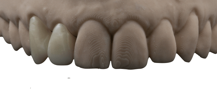
Orthodontic device
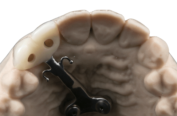
Orthodontic device with skeletal anchorage
Occlusal view
Provisional ortho-prostho device delivery
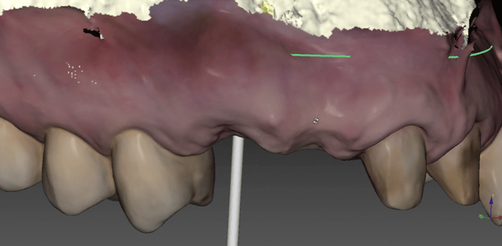
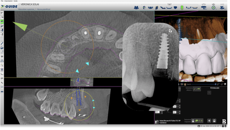
Implant planning at site 1.3; the interdental papilla is visible due to the remaining residual roots.
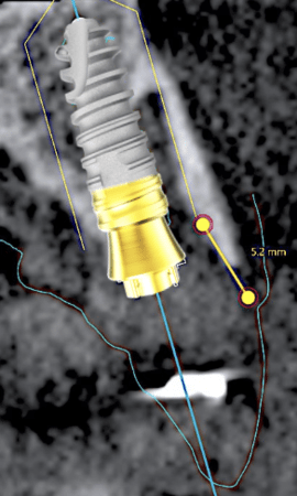
The implant shoulder was planned at least 5 mm apical to the ideal gingival margin at site 1.3.
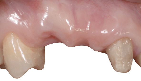
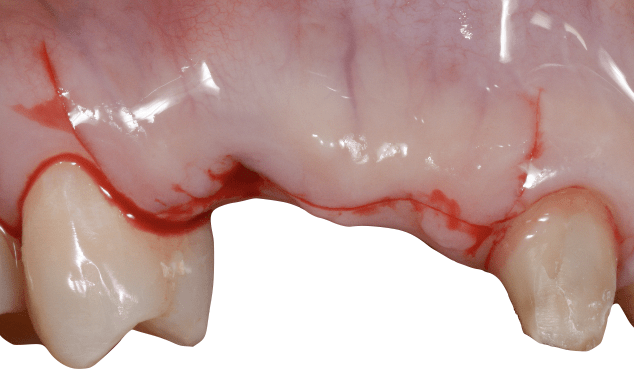
Surgery incision
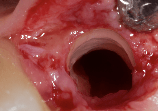
Socket shield 1.3
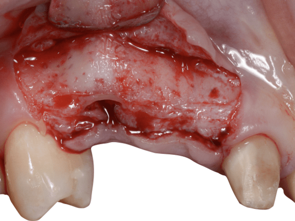
Lateral view of socket shield 1.3
Lateral view of socket shield 1.3
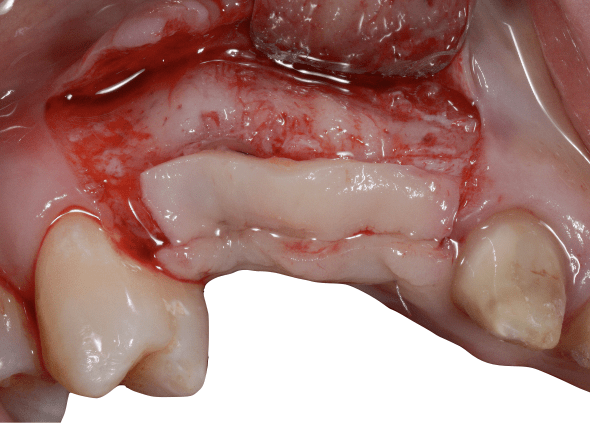
Soft tissue platform technique, CTG graft sutured in occlusal position
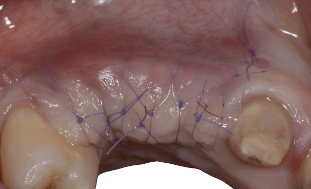
Soft tissue platform technique, suture
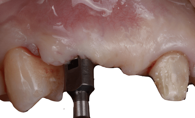
Intermediate abutment connection, bone mill
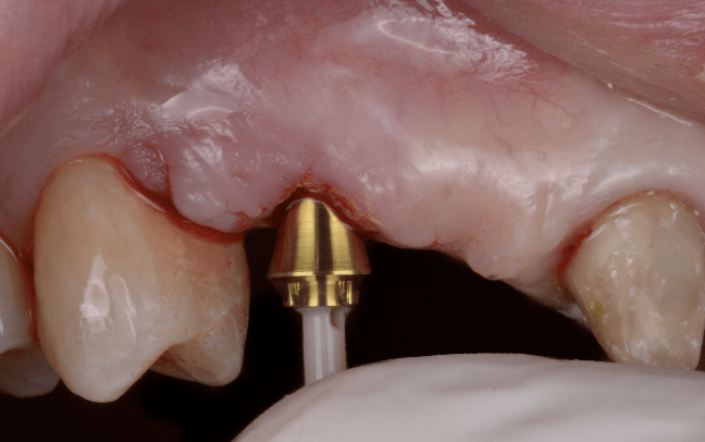
Intermediate abutment connection, N1 base RP 2.5 mm placement
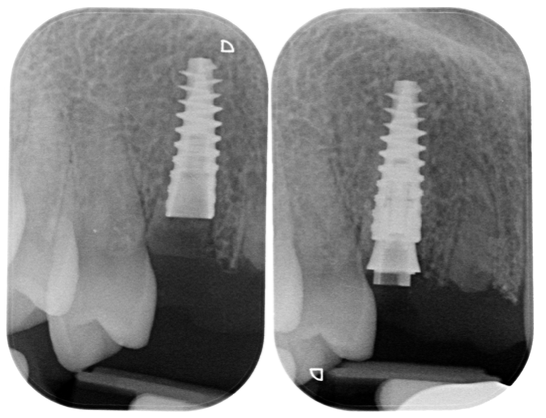
Intermediate abutment connection, N1 base RP 2.5 mm placement
3 months after soft tissue augmentation
Smile in provisional phase
Superimposition between baseline and after orthodontic extrusion/ socket shield/ soft tissue augmentation
Orthodontic extrusion/ socket shield/ soft tissue augmentation
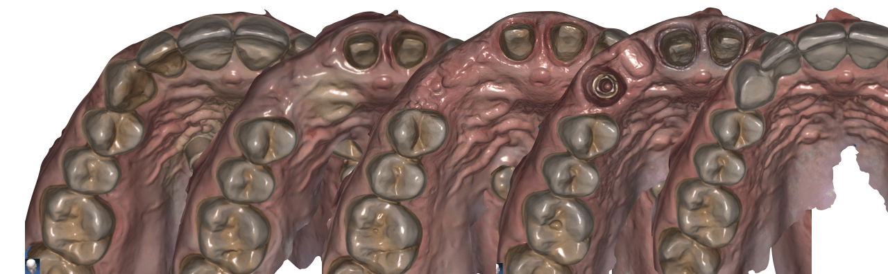
Provisional restorative procedure
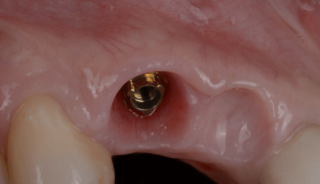
Peri-implant soft tissues before papilla 1.3, 1.2 augmentation surgery
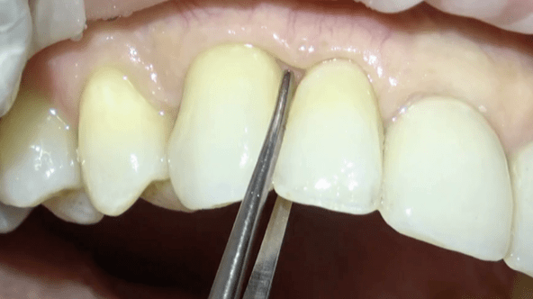
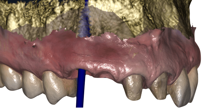
Guided peri-implant papilla 1.3-1.2 augmentation surgery
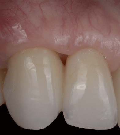
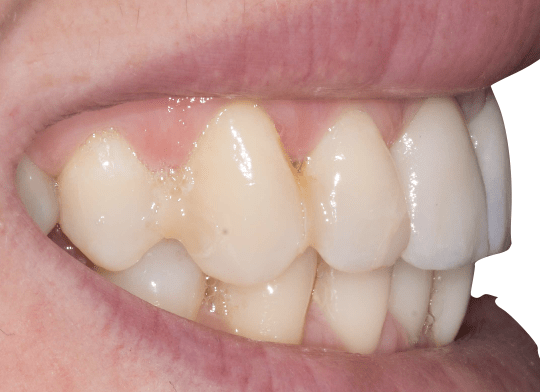
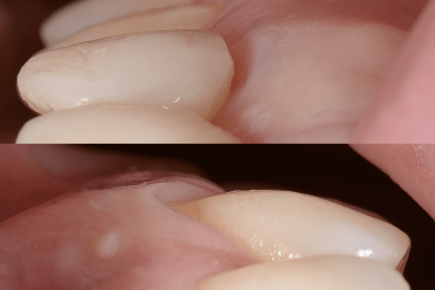
Lateral intraoral view before papilla augmentation surgery
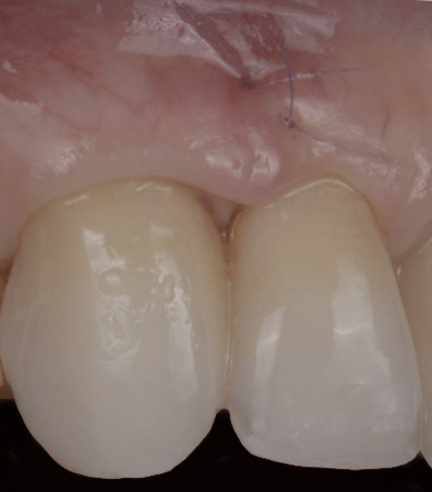
Lateral view intraoral immediately after papilla augmentation surgery
The treatment began with orthodontic extrusion of the roots of teeth 1.3 and 1.2 using skeletal anchorage, aiming to coronally displace hard and soft tissues and improve the biological environment prior to extraction. Following root extrusion, residual fragments were removed and a fixed provisional orthodontic device was delivered to maintain aesthetics and patient comfort during the treatment process.
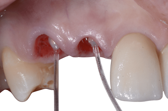
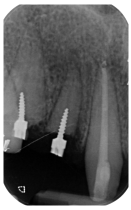
In February 2024, a double socket shield technique was performed, preserving the buccal root fragments of both adjacent teeth. Simultaneously, an implant was placed in the canine site using surgical dynamic navigation, and vertical and horizontal soft tissue augmentation was carried out using the connective tissue platform technique. A second surgical intervention in June 2024 focused on further papilla augmentation, digitally guided to refine soft tissue contours before final prosthetic rehabilitation.
Follow-up & Outcome
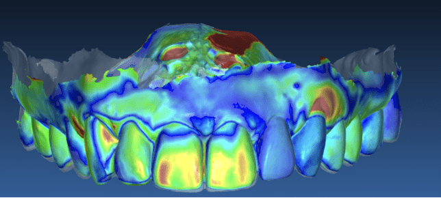
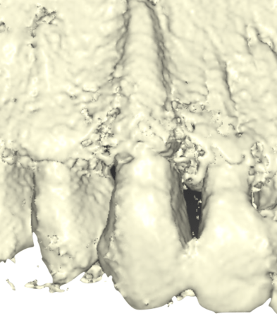
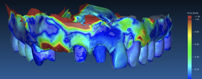
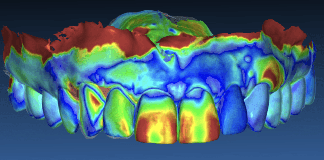
3D colorimetric analysis (superimposition between baseline and after peri-implant papilla augmentation at 1.3 and 1.2)
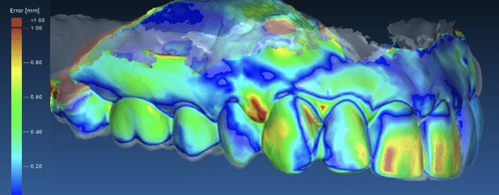
Superimposition between baseline and after peri-implant papilla augmentation 1.3, 1.2.
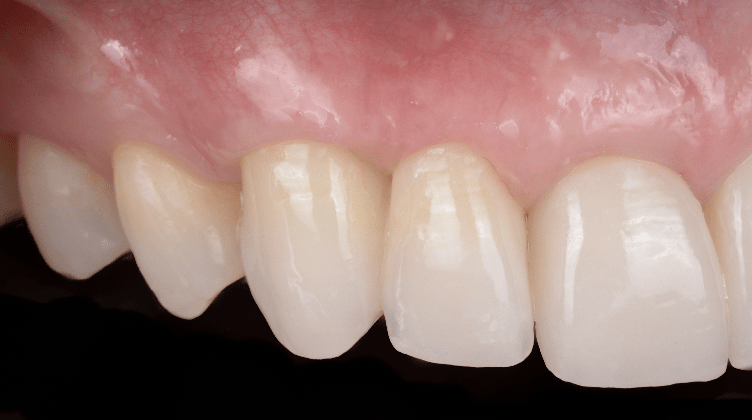
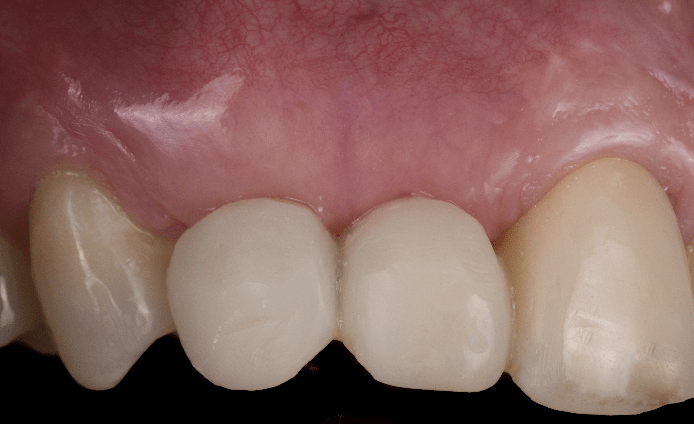
Final outcome, intraoral lateral view
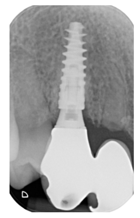
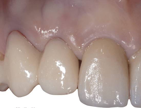
Double socket shields and N1 implant + N1 base + Universal AB base restoration with 1.2 cantilever
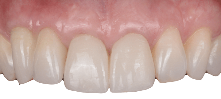
Intraoral frontal view
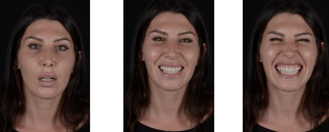
Smile analysis
Clinical follow-up demonstrated stable osseointegration of the implant and favorable soft tissue healing, with harmonious integration into the surrounding natural dentition. The peri-implant papilla showed stable volume and height over time, despite the presence of multiple high-risk aesthetic factors, including replacement of two adjacent teeth in the esthetic zone and a high smile line.
From both functional and aesthetic perspectives, the final outcome met the patient’s expectations, achieving natural tooth proportions, adequate soft tissue volume, and restoration of the canine eminence. This case highlights that, through precise digital planning and the combined use of orthodontic extrusion, socket shield techniques, and guided soft tissue augmentation, highly aesthetic results can be achieved while avoiding bone augmentation and more invasive surgical procedures.