-
0
Thick Phenotypes & Immediate Provisionals
00:00 - 03:00
-
1
Common Mistakes & Lessons Learned
03:00 - 07:00
-
2
The Buccal Pedicle Flap Technique
07:00 - 10:00
-
3
Modified Flap Designs & Collagen Matrix
10:00 - 14:00
-
4
Complex Reconstructions & Soft‑Tissue Stability
14:00 - 18:00
-
5
One‑Step Bone & Soft Tissue, Final Remarks
18:00 - 22:00
- 6 Community questions
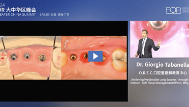
Long‑term success through peri‑implant soft tissue management (Part 2): Boosting the “soft‑tissue collar” for stability
Video highlights
- Key mistakes in peri‑implant tissue management—and how to avoid them
- The buccal pedicle flap and its “dead‑space” concept
- Modified flap designs using collagen matrices
- Achieving stable pink esthetics and long‑term peri‑implant health
- Integrating TiUltra®, Xeal®, and On1™ components to preserve soft‑tissue integrity
In this continuation of his lecture from the FOR Greater China Symposium 2024, Dr. Giorgio Tabanella showcases advanced techniques for “boosting” the peri‑implant mucosa to prevent complications and achieve faster, more predictable esthetic outcomes. Drawing on decades of experience (including his own early failures), he explains how strategic flap designs can minimize repeated surgeries and reduce scar tissue formation.
A highlight of Part 2 is the buccal pedicle flap, a minimally invasive approach that relocates thick palatal tissue to the buccal side—often without needing a free connective‑tissue graft. By creating a deliberate “dead space” or “wrinkle zone” beneath the flap, clinicians can gain significant horizontal thickness, improve the band of keratinized tissue, and even achieve minor vertical augmentation. Dr. Tabanella presents both classic and “modified” flap designs, sometimes in combination with collagen matrices (e.g., Creos Xenoprotect) to further support tissue volume.
Beyond flap design, he emphasizes:
- Preserving at least ~1.4 mm of buccal bone (“buccal bone balcony” or bundle bone) to stabilize the implant site long‑term
- Intrasulcular incisions and sufficient flap release to avoid tension, scar formation, or peri‑implant recession
- Thicker tissue (≥2 mm) around implants to reduce bone loss, food impaction, and inflammation
- Immediate vs. staged loading with careful abutment selection (e.g., TiUltra®, Xeal®, On1™) so that prosthetic changes occur at the sulcus level, preserving the mucosal seal
Clinical cases reveal how small mistakes—like narrow incisions or neglecting frenal attachments—can force multiple re‑entries, added grafts, and months of provisionalization. In contrast, Dr. Tabanella’s one‑step “bone + soft‑tissue” approach (guided bone regeneration plus a buccal pedicle flap) can accelerate healing, simplify the prosthetic phase, and yield a natural, “biomimetic” pink esthetic.
He concludes by highlighting long‑term follow‑ups (e.g., 4–6 years) with stable tissue, well‑filled embrasures, and no significant tissue recession—attributes that mirror healthy gingiva around natural teeth. “Mother nature,” he notes, is often the best ally when clinicians create a favorable environment for the soft tissue to thrive.


