Static Surgical Guide Combined with Dynamic Navigation for Immediate Implant Placement and Provisionalization with AI-Assisted Biomimetic Emergence Profile Reconstruction
Dr. Dingxiang Yuan, an oral surgeon at Beijing Stomatological Hospital, Capital Medical University, specializes in digital technologies for oral aesthetics and functional restoration. As a junior instructor with the Chinese Stomatological Association (CSA), he actively promotes the integration of digital tools in clinical practice. His work has been recognized internationally through presentations at EAO and FDI, and he was awarded second place in the FOR APAC Clinical Case Competition for his innovative, digitally guided implant approach.
From Expertise to Clinical Application
A 28-year-old male patient with no relevant systemic medical history or known drug allergies presented to our department following maxillary anterior dental trauma sustained one month prior. Extraoral and intraoral clinical examination revealed fair oral hygiene with visible calculus deposits and mild generalized gingival inflammation.
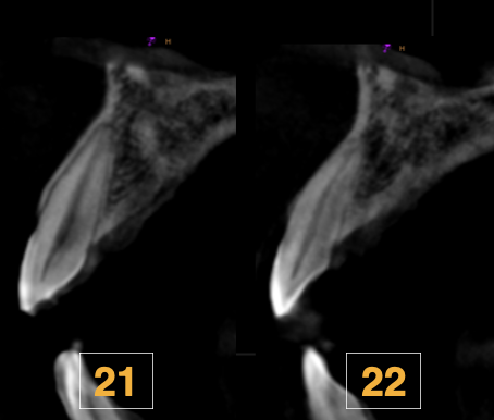
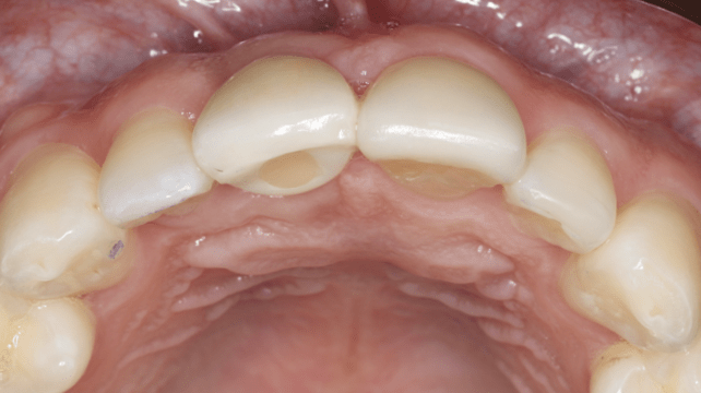
Anterior occlusal analysis demonstrated a deep overbite and deep overjet relationship. Tooth 11 was deemed non-restorable due to its condition as a residual root; notably, the gingival margins and interdental papillae surrounding the affected site were essentially symmetrical with the contralateral anatomy, representing a favorable baseline for soft tissue preservation throughout subsequent treatment. Teeth 21 and 22 presented with incisor crown defects, and the facial soft tissue contour assessed from the occlusal perspective was found to be largely symmetrical to the contralateral reference tooth.
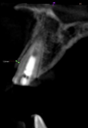
Radiological examination of tooth 11 revealed a facial bone plate thickness of approximately 1 mm, with sufficient bone volume available in the apical region to support implant placement. Based on the comprehensive clinical and radiographic assessment, the following diagnoses were established: residual root at tooth 11, crown defects at teeth 21 and 22, and Stage II periodontitis.
These findings collectively guided the formulation of a multidisciplinary, digitally driven treatment plan aimed at achieving both functional rehabilitation and optimal anterior aesthetic outcomes while preserving the existing soft tissue architecture.
Evaluation & Diagnosis
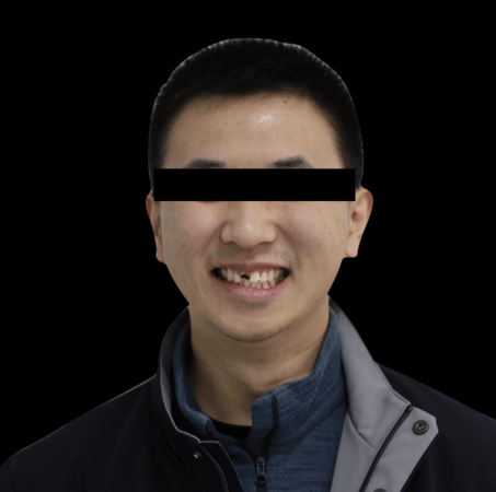
Suffered maxillary anterior teeth trauma a month ago.
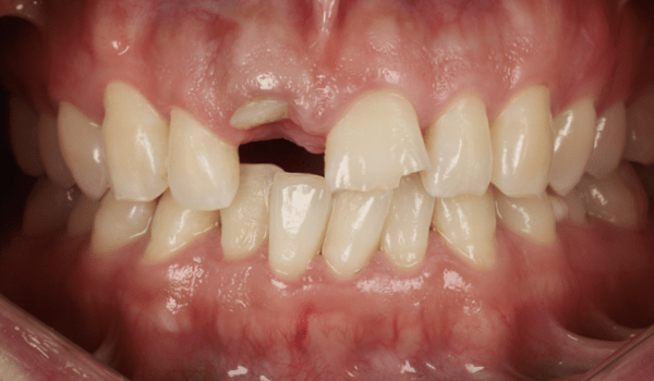
Gingiva exhibited mild inflammation. Anterior teeth occlusion presented as deep overbite and deep overjet.
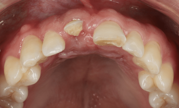
The contour of the facial soft tissue was clinically symmetrical to the contralateral tooth.
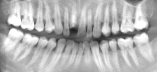
The thickness of the facial bone plate was 1 mm, with sufficient apical bone volume.
Progress & Completion
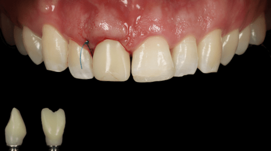
The provisional restoration was delivered out of occlusion and without functional contacts.
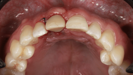
The provisional restoration was delivered out of occlusion and without functional contacts.
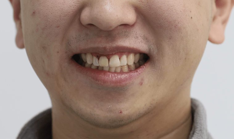
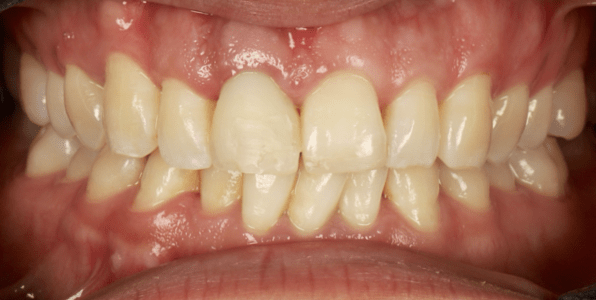
The gingival margin and papilla height were well-maintained.
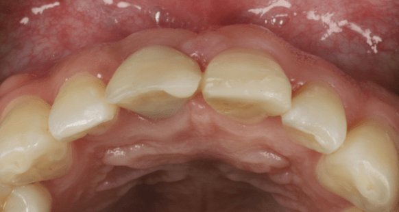
Facial contour of Teeth 11 and 21 were clinically symmetrical.
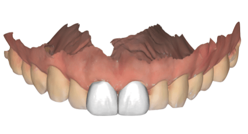
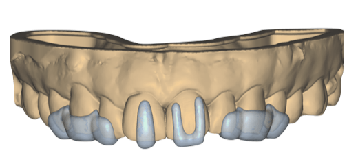
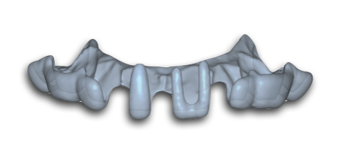
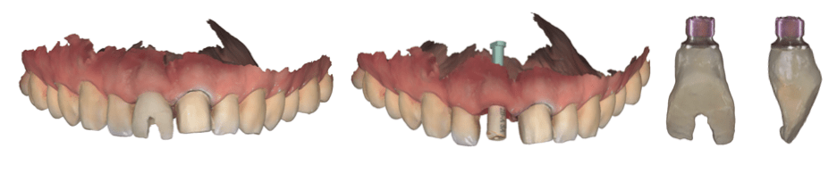
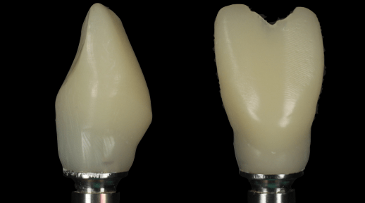
To ensure precise replication of the emergence profile, a triple-scan protocol was followed.
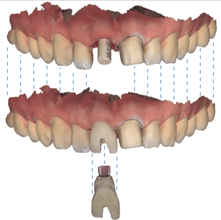
The triple-scan datasets were accurately superimposed based on identical geometric landmarks.
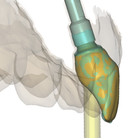
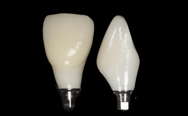
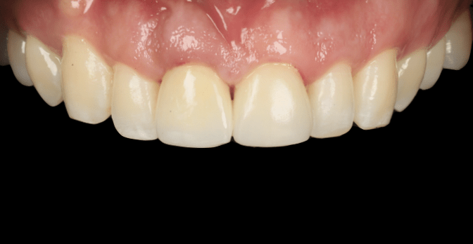
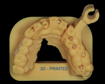
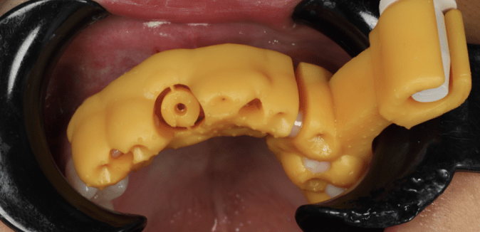
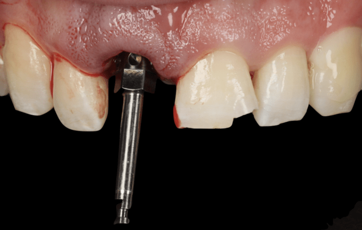
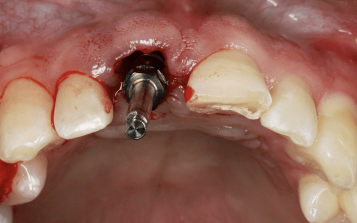
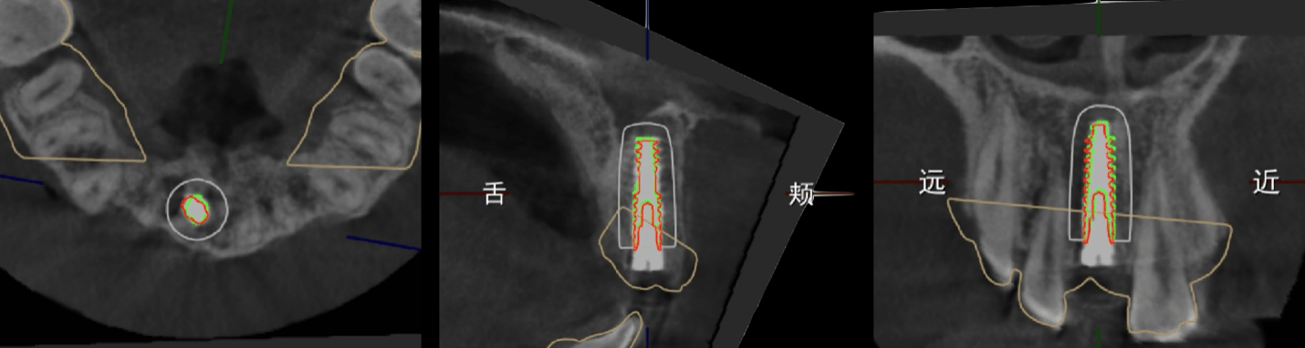
Following periodontal therapy, a fully integrated digital workflow was implemented combining 3D Digital Smile Design, AI-assisted biomimetic emergence profile reconstruction, and a dual static-dynamic surgical guidance system for immediate implant placement at site 11. The AI-extracted transmucosal morphology of the natural tooth served as the biomimetic reference for provisional design, which was fabricated via 3D printing and delivered out of occlusion and without functional contacts to promote optimal peri-implant soft tissue conditioning.
Postoperative accuracy analysis confirmed minimal deviations across all implant position parameters, validating the precision of the combined guidance approach. Dual-Zone Bone Grafting was performed to ensure hard tissue stability, and at the six-month provisionalization checkpoint, gingival margins and papilla height were well maintained, enabling progression to the definitive restorative phase. Final restorations were completed following a triple-scan protocol for precise emergence profile replication, microscopic tooth preparation guided by the preoperative DSD, and delivery of the implant crown at site 11, a veneer on tooth 21, and a composite filling on tooth 22.
Follow-up & Outcome
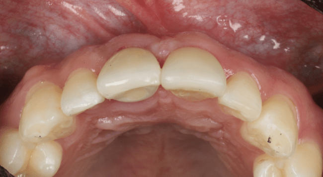
The patient's pink-white aesthetics were well restored.
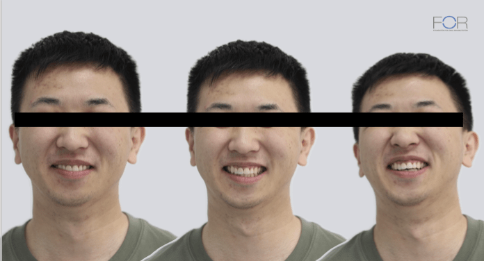
The patient was highly satisfied with the final restoration's aesthetics and function.
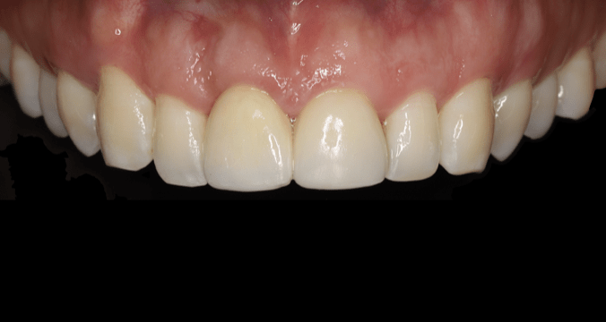
The height of the gingival margin and papillae remained optimal,
Stable maintenance of the soft tissue contour.
At the six-month post-definitive restoration follow-up, the patient demonstrated stable and highly satisfactory pink-white aesthetic outcomes, with gingival margin and papilla heights remaining optimal and the soft tissue contour of teeth 11 and 21 exhibiting near-perfect facial symmetry. The patient reported high satisfaction with both the aesthetics and function of the final restorations.
This case illustrates how the synergistic integration of AI-assisted biomimetic design, combined static-dynamic surgical guidance, and a structured digital prosthetic workflow can reliably achieve precise implant positioning, predictable soft tissue management, and favorable six-month esthetic and functional outcomes in the anterior maxilla.