Immediate implant placement in the molar region with an alternative osteotomy approach
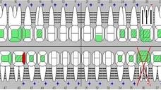
A 25-year-old female patient in good general health condition presented herself for a general dental checkup. The checkup revealed a deep undermining carious lesion on the lingual surface of the endodontically treated tooth #36 (FDI) / #19 (US). The lesion extended apically to the alveolar crest, making it impossible to restore this tooth with a crown. The patient opted for the removal of the hopeless tooth and restoration with an implant-supported crown. The panoramic radiograph displayed a wide interradicular septum allowing for an immediate implant placement procedure using the “pre-extractive interradicular implant bed preparation” technique documented by Rebele et al. This is an alternative osteotomy approach for immediate implant placement in posterior sites.
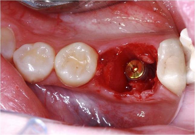
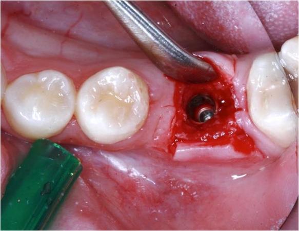
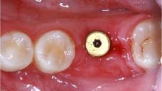
After decoronation and root separation with a diamond bur, the pilot osteotomy and all subsequent osteotomies have been performed through the center of the existing tooth and its roots. Then both roots were removed using a periotome and ensuring a minimally traumatic extraction. A NobelActive WP implant (5,0mm x 11,5mm) was inserted immediately after extraction with an insertion torque of 35 Ncm and closed with a cover screw. Due to the increased incongruency between the alveolus and the much smaller implant diameter, a GBR procedure was performed to help regenerate and close the remaining “gap”. A slowly resorbing anorganic bovine bone mineral (Bio-Oss®, Geistlich Pharma AG, Wolhusen, Switzerland) was applied around the implant into the alveolus and covered by a Creos™ xenoprotect membrane (Nobel Biocare AG, Kloten, Switzerland). The site was then left for open healing. After a healing time of 6 months the second stage surgery was performed, splitting the crestally keratinized soft tissue. A layer of approximately 2mm of newly formated bone and Bio-Oss particles could be observed on top of the implant. After removal of the excess bone a healing abutment with a height and width of 5mm was inserted and left in place for 4 more weeks.
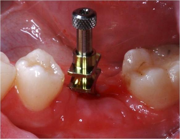
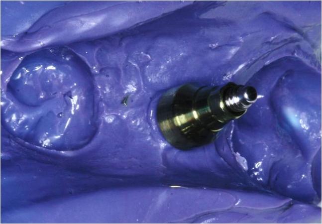
For the prosthetic phase an impression of the implant shoulder with a 5mm impression coping was taken.
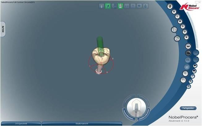
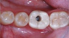
Screw-retained crowns on implants have been documented in literature to provide higher implant survival rates due to the absence of any residual cementation materials. On the other hand there is also documentation in the literature showing higher prosthetic complication rates for screw-reatined crowns, such as chippings due to the position of the screw access hole in the center of the crown. In order to avoid any of these possible complications, both prosthetic and periodontal in nature, a screw-retained full contour zirconia crown (FCZ Crown, Nobel Biocare AG, Kloten, Switzerland) placed on an angulated screw channel abutment (ASC Abutment, Nobel Biocare AG, Kloten, Switzerland) was manufactured in this case. The ASC abutment also presents its advantages in the posterior region especially in patients with a small mouth opening, making it possible to tighten the screw which is angulated towards the mesial.


